
Upper Endoscopy
Upper Endoscopy is a common medical procedure used to examine the upper part of the digestive system. It helps healthcare providers diagnose and sometimes treat conditions affecting the esophagus, stomach, and the first part of the small intestine.
Key Takeaways
- Upper Endoscopy is a procedure to visualize the upper digestive tract using a thin, flexible tube with a camera.
- It is performed to investigate symptoms like persistent heartburn, difficulty swallowing, abdominal pain, and unexplained bleeding.
- Preparation typically involves fasting for several hours before the procedure to ensure a clear view.
- During the procedure, patients are usually sedated, and a scope is gently passed through the mouth into the digestive tract.
- Recovery is generally quick, with most patients returning to normal activities within a day, though some temporary discomfort may occur.
What is Upper Endoscopy?
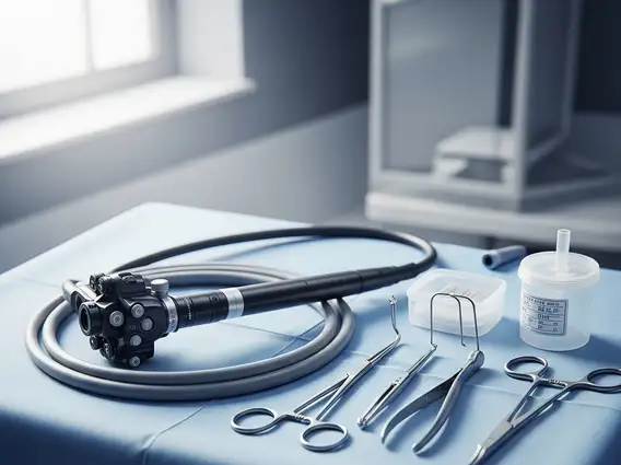
Upper Endoscopy, also known as esophagogastroduodenoscopy (EGD), is a diagnostic and therapeutic medical procedure that allows a doctor to visually examine the lining of the upper part of your gastrointestinal (GI) tract. This includes the esophagus (the tube connecting your mouth to your stomach), the stomach, and the duodenum (the first part of the small intestine). A thin, flexible tube called an endoscope, equipped with a light and a camera, is gently guided through the mouth and down into the GI tract, transmitting images to a video screen. This enables the physician to identify abnormalities, take tissue samples (biopsies), and perform minor interventions.
Reasons for an Upper Endoscopy
There are numerous reasons for upper endoscopy, primarily driven by persistent or concerning symptoms that suggest an issue within the upper digestive system. This procedure is crucial for diagnosing conditions that might not be visible through other imaging techniques. According to the American Society for Gastrointestinal Endoscopy, millions of upper endoscopies are performed annually in the United States to investigate a wide range of gastrointestinal complaints.
Common indications for this procedure include:
- Persistent heartburn or gastroesophageal reflux disease (GERD) symptoms that do not respond to medication.
- Difficulty swallowing (dysphagia) or painful swallowing (odynophagia).
- Unexplained abdominal pain, nausea, vomiting, or persistent indigestion.
- Gastrointestinal bleeding, which may manifest as black, tarry stools (melena) or vomiting blood.
- Anemia due to suspected blood loss from the upper GI tract.
- Monitoring or surveillance for conditions such as Barrett’s esophagus or celiac disease.
- Removal of foreign objects or polyps, and dilation of strictures (narrowings).
Upper Endoscopy Procedure, Preparation, and Recovery
The comprehensive process of an upper endoscopy procedure explained involves several key stages, from initial preparation to post-procedure recovery. Proper adherence to instructions ensures the safety and effectiveness of the examination.
Preparation
Effective upper endoscopy preparation and recovery begins before the procedure itself. Patients are typically instructed to fast for at least six to eight hours prior to the endoscopy, meaning no food or drink (including water). This ensures the stomach is empty, providing a clear view for the physician and minimizing the risk of aspiration. Certain medications, especially blood thinners, may need to be adjusted or temporarily stopped, and patients should discuss their full medication list with their doctor well in advance.
The Procedure
Upon arrival, patients are usually given a sedative intravenously to help them relax and minimize discomfort; a local anesthetic spray may also be applied to the throat to numb it. The patient lies on their side, and a mouth guard is placed to protect the teeth and endoscope. The endoscope is then gently inserted through the mouth, down the esophagus, into the stomach, and finally into the duodenum. Air is sometimes introduced to expand the digestive tract for better visualization. The physician carefully examines the lining, and if necessary, takes biopsies or performs minor therapeutic interventions. The procedure typically lasts between 15 to 30 minutes.
Recovery
After the procedure, patients are monitored in a recovery area until the effects of the sedative wear off. It is common to experience a sore throat, mild bloating, or gas, which usually subside within a few hours. Due to the sedation, patients are not permitted to drive and must arrange for someone to take them home. Most individuals can resume their normal diet and activities the following day, though it’s advised to avoid strenuous activities for the remainder of the day. Any severe or persistent symptoms, such as significant pain, fever, or bleeding, should be reported to the doctor immediately.


