
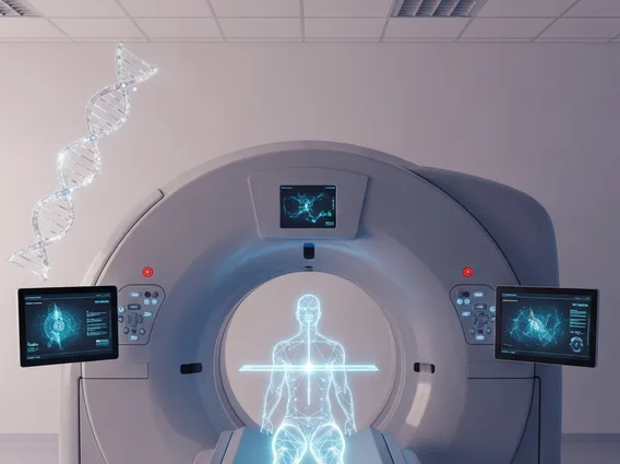
Tomography
Tomography is a sophisticated imaging technique crucial for modern medicine, allowing healthcare professionals to visualize internal structures of the body with remarkable detail. This method provides cross-sectional images that are invaluable for diagnosis, treatment planning, and monitoring various medical conditions.
Key Takeaways
- Tomography creates detailed cross-sectional images of the body’s internal structures.
- It operates by acquiring multiple images from different angles, which are then reconstructed into 3D views.
- Key types include CT, MRI, PET, and SPECT, each utilizing different physical principles.
- Clinical applications range from diagnosing diseases to guiding surgical procedures and monitoring treatment effectiveness.
What is Tomography?
Tomography refers to an advanced medical imaging technique that generates cross-sectional images, or “slices,” of the human body. Unlike traditional X-rays that produce a single, flat image, tomography allows for the visualization of internal structures in three dimensions, providing a clearer and more detailed view. This capability is vital for detecting abnormalities, assessing organ health, and understanding the precise location and extent of various conditions. The process involves capturing multiple images from different angles around the body, which are then computationally processed to reconstruct a comprehensive 3D representation.
How Tomography Works and Its Types
The fundamental principle behind how these imaging techniques function involves acquiring a series of images from various angles around a specific area of the body. These individual images, which are essentially two-dimensional projections, are then fed into powerful computer algorithms. The algorithms process this data to reconstruct detailed cross-sectional slices, effectively eliminating the superimposition of structures that can obscure findings in conventional radiography. This reconstruction allows clinicians to view organs, bones, and soft tissues from multiple perspectives, revealing intricate details that would otherwise be hidden.
There are several distinct types of tomographic scans, each employing different physical principles to generate images:
- Computed Tomography (CT): This method uses X-rays rotated around the patient to create cross-sectional images. CT scans are excellent for visualizing bone structures, detecting internal bleeding, and identifying tumors.
- Magnetic Resonance Imaging (MRI): MRI utilizes strong magnetic fields and radio waves to produce highly detailed images of soft tissues, such as the brain, spinal cord, muscles, and ligaments, without using ionizing radiation.
- Positron Emission Tomography (PET): PET scans use a small amount of radioactive tracer injected into the body to show how organs and tissues are functioning. It is particularly useful in oncology for detecting cancer, assessing its spread, and monitoring treatment response.
- Single-Photon Emission Computed Tomography (SPECT): Similar to PET, SPECT also uses radioactive tracers, but it detects gamma rays. It is often used to analyze the function of internal organs, including the heart, brain, and bones.
Clinical Applications of Tomography
The broad utility of tomography has made it an indispensable tool across numerous medical specialties. Tomography applications span diagnosis, treatment planning, and disease monitoring, significantly enhancing patient care. For instance, in oncology, tomographic imaging is routinely used to detect tumors, determine their size and location, assess their stage, and monitor the effectiveness of chemotherapy or radiation therapy. It allows clinicians to precisely target radiation beams during cancer treatment, minimizing damage to surrounding healthy tissues.
Beyond cancer, tomography plays a critical role in diagnosing cardiovascular diseases by visualizing blood vessels and heart structures, identifying blockages or abnormalities. In neurology, it helps in detecting strokes, brain tumors, and degenerative diseases like Alzheimer’s. Orthopedic specialists use tomography to evaluate complex bone fractures, joint injuries, and spinal conditions, providing detailed anatomical information for surgical planning. Furthermore, in emergency medicine, CT scans are crucial for rapidly assessing internal injuries following trauma, enabling quick and informed decisions that can be life-saving. The ability of tomography to provide non-invasive, detailed views of the body’s interior has revolutionized diagnostic capabilities and continues to improve patient outcomes.


