
Thoracentesis
Thoracentesis is a medical procedure performed to remove excess fluid from the space between the lungs and the chest wall, known as the pleural space. This intervention serves both diagnostic and therapeutic purposes, helping to identify the cause of fluid buildup and alleviate associated symptoms.
Key Takeaways
- Thoracentesis involves the removal of fluid from the pleural space, which surrounds the lungs.
- The procedure is used to diagnose the cause of fluid accumulation (pleural effusion) and to relieve symptoms like shortness of breath.
- It is typically performed under local anesthesia with ultrasound guidance, involving the insertion of a needle into the chest.
- Common risks include pneumothorax (collapsed lung), bleeding, and infection, though serious complications are rare.
- Thoracentesis recovery time is generally quick, with most patients returning to normal activities within a day.
What is Thoracentesis?
Thoracentesis is a medical procedure designed to remove fluid from the pleural space, the narrow area between the visceral and parietal pleura that line the lungs and chest wall, respectively. Normally, this space contains only a small amount of lubricating fluid. However, various medical conditions can lead to an abnormal accumulation of fluid, a condition known as pleural effusion. This excess fluid can compress the lungs, making breathing difficult and causing symptoms such as shortness of breath, chest pain, and coughing.
The primary purposes of thoracentesis are twofold: diagnostic and therapeutic. Diagnostically, the fluid collected during the procedure can be analyzed in a laboratory to determine its composition and identify the underlying cause of the effusion, such as infection, inflammation, cancer, or heart failure. Therapeutically, removing a significant amount of fluid can relieve pressure on the lungs, thereby improving breathing and alleviating discomfort for the patient. According to a study published in the journal Chest, pleural effusions are a common clinical problem, with an estimated 1.5 million cases diagnosed annually in the United States alone, highlighting the importance of procedures like thoracentesis for management and diagnosis.
The Thoracentesis Procedure Explained
The thoracentesis procedure explained involves several key steps to ensure patient safety and effectiveness. Before the procedure, the patient typically undergoes a physical examination and imaging tests, such as a chest X-ray or ultrasound, to confirm the presence and location of the pleural effusion. During the procedure, the patient is usually seated upright, leaning forward over a table, or lying on their side. The physician will then clean and sterilize an area on the back or side of the chest, usually below the shoulder blade.
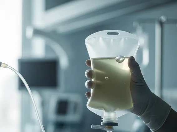
Local anesthetic is injected into the skin and deeper tissues to numb the area, minimizing discomfort. Once the area is numb, a thin needle or catheter is carefully inserted through the chest wall into the pleural space. This step is often guided by ultrasound imaging, which allows the physician to visualize the fluid and avoid vital structures like the lungs or blood vessels. Once the needle is in place, the fluid is slowly withdrawn, either into syringes or a collection bag. The amount of fluid removed depends on the patient’s condition and the purpose of the procedure, but typically ranges from a few hundred milliliters to over a liter. After sufficient fluid has been removed, the needle is withdrawn, and a sterile dressing is applied to the puncture site.
Risks and Recovery After Thoracentesis
While generally safe, there are potential risks of thoracentesis that patients should be aware of. The most common complication is a pneumothorax, or collapsed lung, which occurs if air leaks into the pleural space during or after the procedure. Other potential risks include bleeding, infection at the puncture site, pain or discomfort, and, rarely, injury to nearby organs such as the liver or spleen. Serious complications are infrequent, with studies indicating a pneumothorax rate of approximately 2-6% and a bleeding complication rate of less than 1% for image-guided procedures. Patients are typically monitored for a few hours after the procedure, and a chest X-ray may be performed to check for pneumothorax.
The thoracentesis recovery time is usually quite rapid. Most patients can return home on the same day as the procedure. It is common to experience some mild pain or discomfort at the puncture site for a day or two, which can usually be managed with over-the-counter pain relievers. Patients are generally advised to avoid strenuous activities, heavy lifting, and vigorous exercise for 24 to 48 hours following the procedure. They should also watch for any signs of complications, such as increasing shortness of breath, severe chest pain, fever, redness, or swelling at the puncture site, and contact their doctor if these symptoms occur. Most individuals experience significant relief from their symptoms shortly after the procedure, allowing for improved breathing and comfort.


