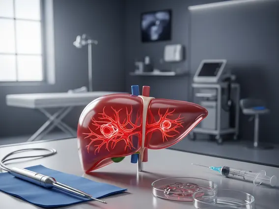
Systematic Biopsy
Systematic biopsy is a critical diagnostic procedure used in medicine to obtain tissue samples from specific areas of an organ, often following a predefined pattern. This method is essential for detecting diseases that may not be visible through imaging alone or that are multifocal in nature.

Key Takeaways
- Systematic Biopsy involves collecting tissue samples from predetermined regions of an organ to diagnose conditions like cancer.
- The procedure often utilizes imaging guidance, such as ultrasound or MRI, to ensure accurate and comprehensive sampling.
- Its primary purpose is to detect abnormalities that might be missed by targeted biopsies, especially in organs where disease can be widespread.
- It is particularly vital in the diagnosis and staging of prostate cancer, where multiple samples are taken from different zones of the gland.
- Pathologists analyze the collected tissue samples to identify cancerous cells or other pathological changes.
What is Systematic Biopsy?
Systematic Biopsy refers to a medical procedure where tissue samples are collected from an organ or area of concern following a predetermined, methodical pattern. Unlike targeted biopsies, which focus on specific lesions identified through imaging, a systematic biopsy aims to comprehensively sample an entire region, even areas that appear normal on scans. This approach is crucial for diagnosing diseases that might be diffusely spread or microscopic, making them difficult to pinpoint with precision. The methodology ensures that a representative collection of tissue is obtained, increasing the likelihood of detecting disease, particularly in early stages. It is a cornerstone diagnostic tool, especially in oncology, where early and accurate detection can significantly impact treatment outcomes.
The Systematic Biopsy Procedure and Its Purpose
The systematic biopsy procedure typically involves the use of imaging guidance, most commonly transrectal ultrasound (TRUS) for prostate biopsies, but sometimes magnetic resonance imaging (MRI) or CT scans for other organs. During the procedure, a thin needle is inserted into the organ, and multiple tissue cores are extracted from various predefined locations. The number of samples and the specific pattern of collection can vary depending on the organ being biopsied and the clinical suspicion. For instance, a common approach might involve taking samples from the anterior, posterior, apex, mid-gland, and base regions of an organ.
The primary purpose of systematic biopsy is to provide a comprehensive histological assessment of the tissue. This method is particularly valuable when a disease, such as cancer, may be present in multiple locations within an organ or when the suspicious area is not clearly defined by imaging. By sampling a wide array of sites, clinicians can gain a more complete picture of the organ’s health, identify the presence of disease, determine its extent, and assess its aggressiveness. This detailed information is vital for accurate diagnosis, staging, and subsequent treatment planning.
Key steps in a typical systematic biopsy procedure include:
- Patient preparation, often including antibiotics to prevent infection.
- Administration of local anesthesia to minimize discomfort.
- Insertion of an imaging probe (e.g., ultrasound) to visualize the organ.
- Systematic collection of multiple tissue cores from predefined zones.
- Labeling and preservation of samples for pathological examination.
Systematic Biopsy for Prostate Cancer Diagnosis
Systematic biopsy for prostate cancer diagnosis is one of its most common and critical applications. Prostate cancer often develops in multiple foci within the prostate gland, and these lesions may not always be visible on standard imaging. Historically, the “sextant” biopsy, involving six samples, was a common systematic approach. However, current guidelines often recommend extended biopsy schemes, which involve taking 10-12 or even more samples from various regions of the prostate, including the peripheral zone, transition zone, and central gland, to improve detection rates. This comprehensive sampling is crucial because approximately 70-80% of prostate cancers originate in the peripheral zone, which is often difficult to assess fully with fewer samples.
According to the American Cancer Society, prostate cancer is the second most common cancer in American men, and biopsy remains the definitive method for diagnosis. While MRI-targeted biopsies are increasingly used for visible lesions, systematic biopsies are often performed concurrently or as a standalone procedure to ensure that clinically significant cancers, particularly those not apparent on MRI, are not missed. The combination of targeted and systematic approaches offers the most thorough evaluation, providing pathologists with sufficient tissue to confirm the presence of cancer, determine its Gleason score (a grading system for prostate cancer), and assess its volume and location, all of which are essential for guiding treatment decisions.