Stage 0 Lip And Oral Cavity Carcinoma In Situ
Stage 0 Lip And Oral Cavity Carcinoma In Situ represents the earliest form of oral cancer, characterized by abnormal cell growth confined to the outermost layer of tissue. Understanding this condition is crucial for early detection and effective intervention, significantly improving patient outcomes.
Key Takeaways
- Stage 0 Lip And Oral Cavity Carcinoma In Situ is the earliest, non-invasive form of oral cancer, where abnormal cells are confined to the surface layer.
- Early detection is vital, as the condition often presents with subtle or no noticeable symptoms.
- Common signs may include persistent white or red patches, or sores that do not heal.
- Treatment primarily involves complete removal of the abnormal tissue, often through surgical excision.
- Regular dental check-ups and self-examinations are crucial for identifying potential signs early.
What is Stage 0 Lip And Oral Cavity Carcinoma In Situ?
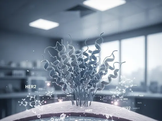
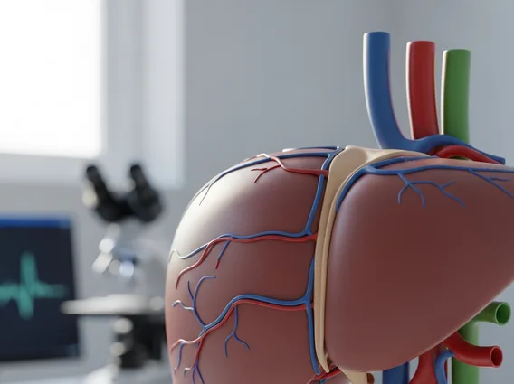
Stage 0 Lip And Oral Cavity Carcinoma In Situ refers to a condition where abnormal, pre-cancerous cells are found only in the outermost layer of the lip or oral cavity lining. This is the earliest stage of oral cancer, meaning the abnormal cells have not yet invaded deeper tissues or spread to other parts of the body. The term carcinoma in situ lip definition specifically highlights that the cancerous cells are “in place” and confined to their original location, making it highly curable if detected and treated promptly. According to the American Cancer Society, early detection of oral and oropharyngeal cancers significantly improves the 5-year survival rate.
This stage is often asymptomatic or presents with very subtle changes, making regular screenings and awareness critical. It signifies a localized cellular change that, if left untreated, has the potential to progress into invasive cancer. The affected areas can include the lips, tongue, gums, inner cheeks, floor of the mouth, and the hard or soft palate. Identifying these changes at Stage 0 is paramount because it allows for less aggressive treatments and a higher probability of complete eradication.
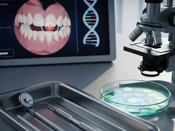
Recognizing Oral Cavity Carcinoma In Situ Symptoms
Recognizing oral cavity carcinoma in situ symptoms can be challenging due to their often subtle nature and resemblance to benign conditions. Unlike more advanced cancers, Stage 0 typically does not involve pain or significant discomfort, which can lead to delayed diagnosis. However, certain persistent changes in the mouth should prompt a medical evaluation.
Potential signs to look for include:
- Leukoplakia: White or grayish patches that cannot be scraped off. These are often found on the tongue, gums, or inside the cheeks.
- Erythroplakia: Red, velvety patches that are slightly raised or flat. Erythroplakia is less common than leukoplakia but has a higher potential for malignancy.
- Non-healing sores: Any sore, ulcer, or lesion on the lip or within the mouth that persists for more than two weeks without healing.
- Unusual lumps or thickenings: Areas of tissue that feel different from the surrounding tissue, even if not painful.
- Persistent irritation: A feeling of roughness, numbness, or a persistent sore throat sensation without an obvious cause.
While these symptoms can be indicative of other, less serious conditions, their persistence warrants immediate medical attention from a dentist or an oral surgeon to rule out carcinoma in situ.
Stage 0 Oral Cancer Treatment Options
The primary goal of stage 0 oral cancer treatment options is the complete removal of the abnormal cells while preserving as much healthy tissue and function as possible. Because the cancer is confined to the superficial layer, treatment is typically highly effective and less invasive compared to later stages.
The most common and effective treatment modality is surgical excision. This involves surgically removing the affected tissue along with a small margin of healthy tissue to ensure all abnormal cells are eliminated. The procedure is often minor and can sometimes be performed under local anesthesia, depending on the size and location of the lesion.
Other treatment approaches may include:
- Laser ablation: Using a focused laser beam to destroy the abnormal cells. This method can be precise and minimize damage to surrounding healthy tissue.
- Cryotherapy: Freezing the abnormal cells to destroy them. This technique is less commonly used for oral carcinoma in situ but can be an option in specific cases.
- Photodynamic therapy (PDT): Involves using a light-sensitive drug and a special light source to kill cancer cells. This is a less common approach for oral carcinoma in situ but may be considered for certain superficial lesions.
Following treatment, regular follow-up appointments are crucial to monitor for recurrence and to detect any new lesions early. Patients are also advised to adopt healthy lifestyle changes, such as quitting smoking and reducing alcohol consumption, to minimize the risk of developing new oral cancers.