Spiral Ct Scan
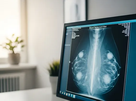
A Spiral CT Scan, also known as helical CT, is an advanced medical imaging technique that uses X-rays to create detailed cross-sectional images of the body. This diagnostic tool is crucial for detecting a wide range of medical conditions, from internal injuries to complex diseases.
Key Takeaways
- Spiral CT Scan is an advanced imaging method that captures continuous, detailed X-ray images.
- It works by rotating X-ray tubes and detectors around the patient while the table moves, creating a helical data path.
- This technology offers significant benefits, including faster scan times, reduced motion artifacts, and improved 3D reconstruction capabilities.
- Common applications include diagnosing cancers, vascular diseases, and internal injuries.
- Key considerations involve radiation exposure and potential reactions to contrast agents.
What is a Spiral CT Scan?
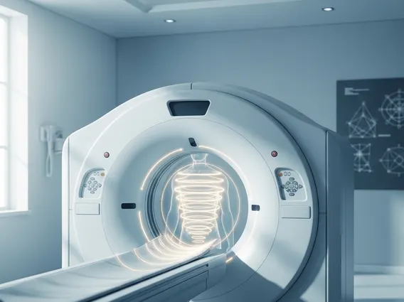
A Spiral CT Scan is a sophisticated medical imaging procedure that utilizes computed tomography (CT) technology to acquire images in a continuous, helical (spiral) path. Unlike conventional CT scans that take images slice by slice with pauses in between, a spiral CT scanner continuously rotates its X-ray tube and detectors around the patient while the examination table simultaneously moves through the gantry. This continuous motion allows for faster data acquisition and the creation of a seamless volume of data, which can then be reconstructed into detailed two-dimensional and three-dimensional images of organs, soft tissues, bone, and blood vessels.
This method significantly reduces scan times and minimizes motion artifacts, making it particularly effective for imaging areas affected by patient movement, such as the chest during breathing. The ability to acquire data volumetrically also enhances the diagnostic quality, providing clinicians with comprehensive views necessary for accurate diagnosis and treatment planning.
How a Spiral CT Scan Works and Its Clinical Applications
The operational principle behind how a spiral CT scan works involves a synchronized movement of the X-ray source and the patient. As the X-ray tube and detectors rotate around the patient, the patient’s bed steadily advances through the scanner’s opening. This coordinated motion generates a continuous, helical projection of X-ray data. Sophisticated computer algorithms then process this raw data to reconstruct detailed cross-sectional images, which can be viewed from various angles or rendered into three-dimensional models. This volumetric data acquisition is particularly beneficial for creating high-resolution images of complex anatomical structures.
The spiral CT scan uses and benefits are extensive across various medical specialties. Its speed and precision make it invaluable for:
- Oncology: Detecting and staging cancers, evaluating tumor response to treatment, and guiding biopsies.
- Vascular Imaging: Identifying aneurysms, blockages, and other vascular abnormalities, often with the aid of contrast agents (CT angiography).
- Trauma Assessment: Rapidly assessing internal injuries in emergency situations, such as those affecting the brain, chest, or abdomen.
- Pulmonary Studies: Diagnosing lung diseases like pulmonary embolism, pneumonia, and interstitial lung disease.
- Musculoskeletal Imaging: Detailed visualization of complex fractures, joint disorders, and spinal conditions.
According to the American College of Radiology, CT scans, including spiral CT, play a critical role in modern medicine, with millions performed annually in the United States, significantly contributing to early disease detection and improved patient outcomes.
Risks and Considerations of Spiral CT Scans
While a powerful diagnostic tool, there are certain risks of spiral CT scan procedures that patients and clinicians must consider. The primary concern is exposure to ionizing radiation. Although the radiation dose from a single CT scan is generally low, cumulative exposure from multiple scans over time can slightly increase the lifetime risk of cancer. Medical professionals carefully weigh the diagnostic benefits against these potential risks, especially in pediatric patients or those requiring repeated imaging.
Another consideration involves the use of intravenous contrast agents, which are sometimes administered to enhance the visibility of blood vessels and certain tissues. While generally safe, these agents can cause allergic reactions in some individuals, ranging from mild symptoms like hives to severe anaphylaxis. Patients with kidney impairment may also be at risk of contrast-induced nephropathy. It is crucial for patients to inform their healthcare provider about any allergies, existing medical conditions, or medications prior to the scan.
Pregnant women are typically advised to avoid CT scans unless absolutely necessary, due to the potential risks of radiation to the developing fetus. In such cases, alternative imaging modalities like ultrasound or MRI may be considered. Open communication with the medical team ensures that the most appropriate and safest imaging strategy is chosen for each patient’s unique circumstances.