Sonogram
A sonogram, also known as an ultrasound, is a non-invasive medical imaging technique that utilizes high-frequency sound waves to create real-time images of structures inside the body. It is a widely used diagnostic tool across various medical specialties.
Key Takeaways
- A Sonogram uses high-frequency sound waves to generate real-time images of internal body structures.
- The technology involves a transducer emitting sound waves and capturing echoes, which are then converted into visual images.
- Sonograms are versatile, employed in obstetrics, cardiology, abdominal imaging, and vascular studies.
- They are a safe and effective diagnostic tool, avoiding ionizing radiation.
- Different types of sonogram procedures are tailored to specific anatomical regions and diagnostic needs.
What is a Sonogram?
A Sonogram is a medical imaging technique that employs sound waves to produce pictures of organs, tissues, and blood flow inside the body. Unlike X-rays, it does not use ionizing radiation, making it a safe option for many diagnostic purposes, including during pregnancy. This process is often referred to as sonogram explained medical imaging, highlighting its role in visualizing internal anatomy without invasive procedures. The images, called sonograms, provide valuable information for diagnosing and monitoring a wide range of medical conditions.
The principle behind a Sonogram involves the emission of sound waves at frequencies beyond the range of human hearing. These waves travel through the body and bounce off structures like organs and blood vessels. The echoes that return are then detected by the same device, allowing a computer to construct a dynamic, real-time visual representation.
How Sonogram Technology Works
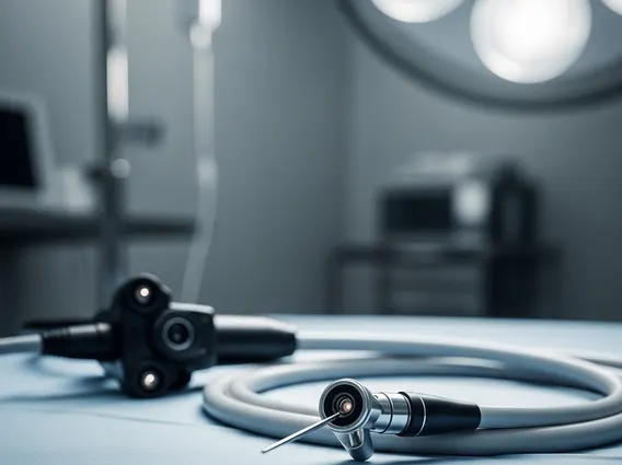
The fundamental principle of how Sonogram technology works relies on the physics of sound waves. A specialized device called a transducer is placed on the skin, often with a gel to ensure good contact and eliminate air pockets. This transducer emits high-frequency sound waves into the body. As these sound waves encounter different tissues and structures, they reflect, or “echo,” back to the transducer.
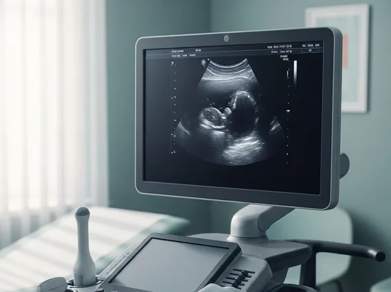
The transducer then detects these returning echoes. The time it takes for the sound waves to travel to a structure and return, along with the intensity of the echoes, provides information about the depth, size, and composition of the tissues. A computer processes this data rapidly, converting it into a two-dimensional image displayed on a monitor. Modern Sonogram systems can also create three-dimensional (3D) and four-dimensional (4D) images, offering more detailed views, particularly useful in obstetrics for visualizing fetal development. According to the World Health Organization (WHO), ultrasound imaging is a cornerstone of medical diagnostics globally, especially in low-resource settings due to its portability and cost-effectiveness.
Applications and Types of Sonogram Procedures
Sonograms are incredibly versatile diagnostic tools, and understanding what a Sonogram is used for reveals its broad utility across medical specialties. They are instrumental in visualizing soft tissues that do not show up well on X-rays, providing critical insights into various conditions. The different types of Sonogram procedures are tailored to specific anatomical regions and diagnostic objectives.
Common applications include:
- Obstetric Sonogram: Used to monitor fetal growth and development, assess gestational age, detect potential abnormalities, and determine the position of the fetus.
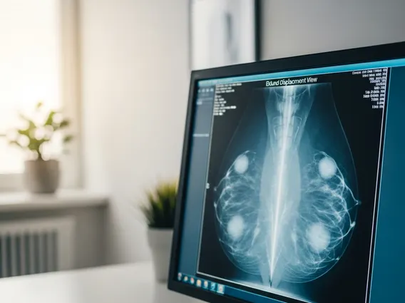
- Abdominal Sonogram: Examines organs such as the liver, gallbladder, pancreas, spleen, and kidneys to diagnose conditions like gallstones, kidney stones, tumors, or fluid collections.
- Cardiac Sonogram (Echocardiogram): Visualizes the heart’s structure and function, assessing blood flow, valve health, and the pumping efficiency of the heart.
- Vascular Sonogram: Evaluates blood vessels to detect blockages, clots, or aneurysms, commonly performed on arteries and veins in the legs, neck, and abdomen.
- Thyroid Sonogram: Used to examine the thyroid gland for nodules, cysts, or other abnormalities.
- Musculoskeletal Sonogram: Assesses muscles, tendons, ligaments, and joints for injuries, inflammation, or other conditions.
Each type of Sonogram procedure requires specific patient preparation and transducer placement to optimize image quality for the targeted area, ensuring accurate diagnosis and effective patient management.