Psma Pet Scan
The PSMA PET Scan represents a significant advancement in medical imaging, particularly in the field of oncology. This sophisticated diagnostic tool offers a highly sensitive method for detecting and staging prostate cancer, providing crucial information for patient management.
Key Takeaways
- A PSMA PET Scan is an advanced imaging technique primarily used for diagnosing and staging prostate cancer.
- It works by detecting the Prostate-Specific Membrane Antigen (PSMA) protein, which is often overexpressed on prostate cancer cells.
- The procedure involves injecting a radioactive tracer that binds to PSMA, followed by a PET scan to visualize cancer cells.
- This scan is highly effective in initial staging, detecting recurrence, and guiding treatment decisions for prostate cancer patients.
What is a PSMA PET Scan?
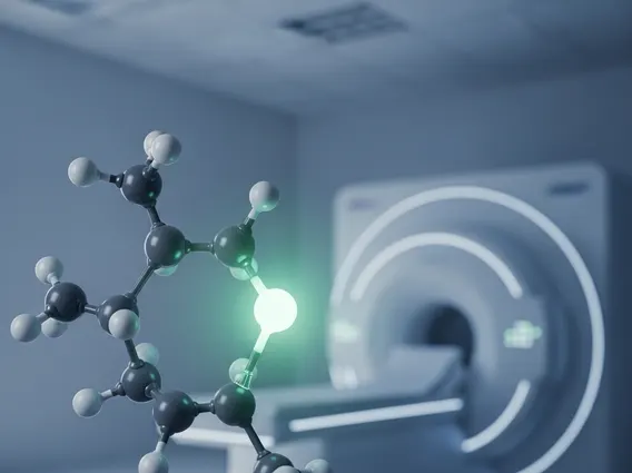
A PSMA PET Scan, or Prostate-Specific Membrane Antigen Positron Emission Tomography Scan, is a specialized imaging test used to detect prostate cancer cells throughout the body. Unlike conventional imaging methods that may struggle to identify small or distant metastases, PSMA PET offers enhanced precision by targeting a specific protein.
This diagnostic technique works by utilizing a small amount of a radioactive tracer that is specifically designed to bind to the PSMA protein. Prostate-Specific Membrane Antigen (PSMA) is a protein found on the surface of prostate cancer cells, often in higher concentrations than on healthy cells. When the tracer is injected into the patient’s bloodstream, it travels throughout the body and attaches to these PSMA-expressing cancer cells. A Positron Emission Tomography (PET) scanner then detects the emissions from the radioactive tracer, creating detailed images that highlight the location and extent of the cancer. This mechanism explains how PSMA PET Scan works, providing clinicians with a clearer picture of the disease’s spread.
PSMA PET Scan Procedure: What to Expect
Undergoing a PSMA PET Scan involves several steps designed to ensure accurate results and patient comfort. Patients are typically advised to fast for a few hours before the scan and to stay well-hydrated. Upon arrival at the imaging center, a small amount of the radioactive tracer is injected, usually into a vein in the arm. This tracer is safe and generally well-tolerated, with minimal side effects.
After the injection, there is a waiting period, typically 60 to 90 minutes, allowing the tracer to circulate throughout the body and accumulate in any cancer cells that express PSMA. During this time, patients are usually asked to rest quietly. Following the uptake period, the patient will lie on a table that slides into the PET scanner. The scan itself usually takes between 15 and 30 minutes, during which it is important to remain as still as possible to ensure clear images. The entire PSMA PET Scan procedure explained involves careful preparation, tracer administration, an uptake period, and the actual scanning process, after which patients can typically resume their normal activities.
- Preparation: Fasting for a few hours and ensuring adequate hydration.
- Tracer Injection: A small amount of radioactive tracer administered intravenously.
- Uptake Period: Resting for 60-90 minutes while the tracer distributes in the body.
- Scanning: Lying still in the PET scanner for 15-30 minutes.
- Post-Scan: Resuming normal activities; drinking fluids to help excrete the tracer.
Role of PSMA PET in Prostate Cancer Detection
The PSMA PET Scan for prostate cancer has revolutionized the way this disease is detected, staged, and monitored. Prostate cancer is a significant health concern globally; according to the World Health Organization (WHO), it is the second most common cancer in men, with an estimated 1.4 million new cases diagnosed worldwide in 2020. This advanced imaging technique offers superior sensitivity and specificity compared to traditional imaging modalities, especially in identifying small lesions or metastases that might otherwise be missed.
PSMA PET scans are particularly valuable in several clinical scenarios. They are used for initial staging in men with high-risk prostate cancer to determine if the cancer has spread beyond the prostate gland. Furthermore, they play a critical role in detecting recurrence in patients who have undergone treatment (such as surgery or radiation) and subsequently experience a rise in their Prostate-Specific Antigen (PSA) levels, indicating potential cancer return. By precisely locating recurrent disease, PSMA PET helps guide salvage therapies, ensuring more targeted and effective treatment strategies.