
Psammoma Body
A Psammoma Body is a microscopic, concentrically laminated, calcified structure found in various tissues, often indicative of certain pathological processes. These unique formations are particularly significant in the diagnosis and understanding of specific benign and malignant conditions.
Key Takeaways
- Psammoma bodies are small, spherical, calcified structures with a characteristic layered appearance.
- Their formation often involves cellular necrosis, dystrophic calcification, and collagen deposition.
- They serve as important diagnostic markers, especially in certain types of cancer.
- Commonly associated conditions include papillary thyroid carcinoma, serous ovarian carcinoma, and meningiomas.
- Understanding their presence aids pathologists in accurate disease classification and prognosis.
What is a Psammoma Body? Definition and Examples
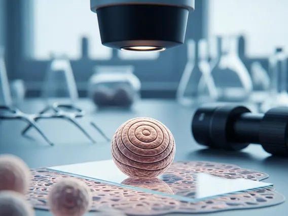
A Psammoma Body refers to a small, round, or oval collection of calcium salts that exhibits a distinctive lamellated, or layered, appearance, resembling tiny grains of sand (from the Greek “psammos” meaning sand). These structures are typically acellular, meaning they do not contain living cells, and range in size from a few micrometers to several millimeters. Their concentric rings are formed by the deposition of calcium and other minerals around a central core, which can be a degenerated cell, a blood clot, or a small piece of necrotic tissue.
The presence of psammoma bodies is a significant finding in histopathology, often pointing towards specific disease entities. For instance, they are classic features in certain tumors, where their identification can be crucial for diagnosis. Examples include their frequent detection in the papillary architecture of papillary thyroid carcinoma and in the stromal elements of serous ovarian carcinomas, providing a key morphological clue for pathologists.
Psammoma Body Formation and Clinical Significance
The psammoma body formation and significance are complex processes involving a sequence of cellular and molecular events. While the exact mechanism can vary depending on the tissue and underlying condition, a common pathway involves the death of cells (necrosis), followed by the calcification of the necrotic debris. This process, known as dystrophic calcification, occurs in damaged or degenerated tissues in the absence of systemic calcium imbalance. The layers are thought to build up as calcium salts are deposited concentrically around a nidus, often a degenerating cell or a small vessel.
From a clinical perspective, the clinical significance of psammoma bodies is primarily diagnostic. Their presence is often highly characteristic of specific neoplastic conditions, making them valuable markers for pathologists. For example, in the context of ovarian tumors, the detection of psammoma bodies strongly suggests a serous subtype, which influences treatment decisions and prognostic assessment. Similarly, their identification in thyroid biopsies is a hallmark of papillary thyroid carcinoma, guiding further diagnostic and therapeutic interventions.
Key aspects of their formation and significance include:
- Necrotic Core: Often originates from a degenerated cell or a small thrombus.
- Dystrophic Calcification: Calcium deposition in damaged tissues.
- Concentric Layering: Gradual accumulation of mineral salts and collagen fibers.
- Diagnostic Marker: Highly indicative of specific tumor types.
- Prognostic Indicator: Can sometimes correlate with tumor behavior or differentiation.
Conditions Associated with Psammoma Bodies
The identification of psammoma body associated conditions is a critical aspect of histopathological diagnosis. These structures are not found universally but are strongly linked to a specific set of benign and malignant pathologies. Their presence often guides the pathologist toward a definitive diagnosis, particularly in oncology.
The most notable conditions where psammoma bodies are frequently observed include:
- Papillary Thyroid Carcinoma: This is one of the most common thyroid cancers, and psammoma bodies are a classic histological feature, often found within the papillary fronds.
- Serous Ovarian Carcinoma: Both low-grade and high-grade serous carcinomas of the ovary, fallopian tube, and peritoneum frequently contain psammoma bodies, particularly within the papillary structures or stroma.
- Meningioma: These are typically benign tumors of the meninges (membranes surrounding the brain and spinal cord). Psammoma bodies are common in psammomatous meningiomas, a specific subtype.
- Endometrial Adenocarcinoma: Less commonly, psammoma bodies can be found in certain subtypes of endometrial cancer.
- Renal Cell Carcinoma: Rarely, they may be seen in some variants of kidney cancer.
- Mesothelioma: In some cases, particularly in papillary forms of peritoneal mesothelioma, psammoma bodies can be present.
While primarily associated with these neoplastic conditions, psammoma bodies can occasionally be found in benign lesions or inflammatory processes, though this is less common and usually requires careful differential diagnosis by a trained pathologist.


