Positron Emission Tomography Scan
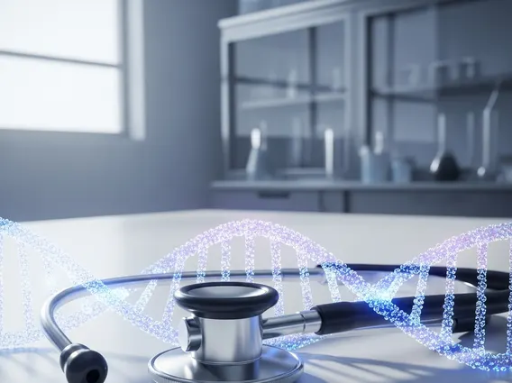
A Positron Emission Tomography Scan, commonly known as a PET scan, is a powerful diagnostic imaging technique used in medicine to visualize metabolic processes in the body. It plays a crucial role in detecting and monitoring various diseases at a cellular level, often before structural changes are visible on other imaging tests.
Key Takeaways
- Positron Emission Tomography Scan (PET scan) is an advanced imaging technique that visualizes metabolic activity in the body.
- It works by detecting gamma rays emitted from a radioactive tracer, which highlights areas of increased cellular activity.
- PET scans are widely used in oncology for cancer detection, staging, and monitoring treatment response.
- They also have significant applications in cardiology and neurology for assessing heart function and brain disorders.
- While generally safe, PET scans involve minimal radiation exposure and potential allergic reactions to the tracer.
What is Positron Emission Tomography (PET) Scan?
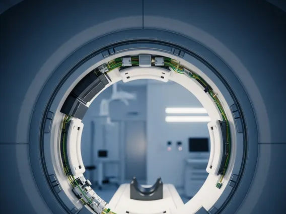
A Positron Emission Tomography Scan (PET scan) is a non-invasive medical imaging test that uses a small amount of a radioactive tracer to produce detailed three-dimensional images of organs and tissues within the body. Unlike traditional imaging methods like X-rays or CT scans that show anatomical structures, a PET scan reveals how well organs and tissues are functioning by measuring metabolic activity. This capability allows clinicians to identify disease processes at their earliest stages, often before they manifest as physical changes detectable by other imaging modalities. The tracer, typically a glucose analog like fluorodeoxyglucose (FDG), accumulates in cells with high metabolic rates, such as cancer cells, making them visible on the scan.
How Does a PET Scan Work?
The operational principle behind a Positron Emission Tomography Scan involves the administration of a radiotracer and the subsequent detection of its emissions. First, a small dose of a radioactive tracer, such as fluorodeoxyglucose (FDG), is injected into the patient’s bloodstream. This tracer travels through the body and is absorbed by cells that are metabolically active. For instance, cancer cells often consume glucose at a much higher rate than healthy cells, causing the FDG to accumulate more intensely in cancerous regions.
Once the tracer is absorbed, it undergoes radioactive decay, emitting tiny particles called positrons. These positrons quickly collide with electrons in the body, resulting in an annihilation event that produces two gamma rays traveling in opposite directions. The PET scanner detects these gamma rays simultaneously. A sophisticated computer then processes this data to reconstruct detailed images that show the distribution and concentration of the tracer throughout the body. Areas with higher tracer uptake appear brighter on the scan, indicating increased metabolic activity, which can signify disease.
Clinical Uses, Benefits, and Risks of PET Scans
Positron Emission Tomography Scans offer significant advantages in modern medicine, providing unique insights into disease processes. The primary clinical applications of a PET scan are diverse, ranging from oncology to neurology and cardiology. Understanding the uses of positron emission tomography scans, as well as their benefits and risks of pet scans, is essential for patients and healthcare providers.
Key clinical uses include:
- Oncology: Detecting cancer, determining its stage, assessing whether cancer has spread (metastasis), evaluating the effectiveness of cancer treatment, and checking for cancer recurrence. According to the American Cancer Society, PET scans are particularly valuable for cancers like lung, colorectal, melanoma, lymphoma, and head and neck cancers.
- Cardiology: Assessing blood flow to the heart muscle, identifying areas of damaged heart tissue, and evaluating myocardial viability after a heart attack.
- Neurology: Diagnosing brain disorders such as Alzheimer’s disease by detecting early changes in brain metabolism, locating seizure foci in epilepsy, and evaluating brain tumors.
The benefits of Positron Emission Tomography Scans include their ability to detect diseases earlier than many other imaging tests, provide functional information about organs, and guide treatment plans more effectively. However, like all medical procedures, there are some risks. These typically involve minimal exposure to ionizing radiation from the radiotracer, which is generally considered safe and quickly eliminated from the body. Rarely, patients may experience an allergic reaction to the tracer or discomfort from the injection. Clinicians carefully weigh these benefits against the potential risks for each patient.