Positron Emission Tomography Computed Tomography Scan
A Positron Emission Tomography Computed Tomography Scan is a sophisticated medical imaging technique that provides detailed insights into the body’s metabolic activity and anatomical structures. This powerful diagnostic tool plays a crucial role in modern medicine, particularly in the fields of oncology, neurology, and cardiology.
Key Takeaways
- A Positron Emission Tomography Computed Tomography (PET/CT) scan combines functional (PET) and anatomical (CT) imaging for comprehensive diagnostic information.
- It utilizes a radioactive tracer, often a glucose analog, to highlight areas of increased metabolic activity, such as tumors.
- The scan is vital for diagnosing, staging, and monitoring various diseases, especially cancers, and assessing treatment effectiveness.
- Patients typically undergo preparation like fasting, receive an intravenous tracer, and then lie still during the combined PET and CT scanning.
- The procedure is non-invasive and provides critical data that helps clinicians make informed decisions about patient care.
What is a Positron Emission Tomography Computed Tomography (PET/CT) Scan?
A Positron Emission Tomography Computed Tomography (PET/CT) scan is an advanced diagnostic imaging technique that merges two distinct imaging modalities into a single examination. Positron Emission Tomography (PET) provides functional information by detecting metabolic changes at the cellular level, while Computed Tomography (CT) offers detailed anatomical images of organs and tissues. By combining these, a PET/CT scan creates highly precise images that reveal both the location and metabolic activity of cells, offering a comprehensive view of the body’s internal state. This integration allows clinicians to pinpoint abnormalities with greater accuracy than either scan could achieve alone.
The core principle involves injecting a small amount of a radioactive tracer, often a glucose analog like fluorodeoxyglucose (FDG), into the patient’s bloodstream. Cells with higher metabolic rates, such as cancer cells, tend to absorb more of this tracer. The PET scanner then detects the gamma rays emitted by the tracer as it decays, creating a map of metabolic activity. Simultaneously, the CT component provides high-resolution anatomical images, which are then fused with the PET data to produce a single, detailed image. This synergistic approach is invaluable for early disease detection and precise localization.
How a PET/CT Scan Works and What to Expect During the Procedure
A PET CT scan procedure explained involves several key stages designed to ensure accurate imaging. The process begins with patient preparation, which typically includes fasting for several hours before the scan to ensure optimal tracer uptake and reduce background glucose levels. Patients are also advised to stay hydrated and avoid strenuous activity prior to the appointment.
Upon arrival, a small amount of a radioactive tracer, most commonly Fluorodeoxyglucose (FDG), is administered intravenously. This tracer is designed to mimic glucose, which is readily absorbed by cells that are metabolically active. After the injection, there is a waiting period, usually 60 to 90 minutes, allowing the tracer to circulate throughout the body and accumulate in target tissues. During this time, patients are asked to rest quietly to minimize muscle activity, which could otherwise absorb the tracer and interfere with image quality.
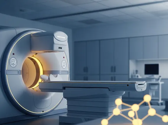
The actual scanning process involves the patient lying still on a comfortable table that slides into the PET/CT scanner. The scanner simultaneously acquires both PET and CT images. The PET component detects the positrons emitted by the tracer, while the CT component captures X-ray images of the body’s internal structures. The combined scan typically takes between 20 to 45 minutes, depending on the area being examined. Throughout the procedure, medical staff monitor the patient and provide instructions. After the scan, patients are generally free to resume normal activities, with advice to drink plenty of fluids to help flush the remaining tracer from their system. The radiation exposure from a PET/CT scan is considered safe and is carefully managed by medical professionals.
Key Clinical Applications of PET/CT Scans
The uses of PET CT scan are extensive and continue to expand across various medical disciplines, with oncology being its most prominent application. In cancer care, PET/CT scans are indispensable for:
- Diagnosis and Staging: Identifying cancerous lesions, determining the extent of the disease (staging), and detecting metastasis (spread to other parts of the body). For instance, PET/CT can detect smaller lesions that might be missed by other imaging techniques.
- Treatment Planning and Monitoring: Guiding biopsies, planning radiation therapy, and assessing the effectiveness of chemotherapy or other treatments by observing changes in metabolic activity. A reduction in tracer uptake often indicates a positive response to therapy.
- Recurrence Detection: Monitoring for cancer recurrence after treatment, often before structural changes are visible on conventional imaging.
Beyond oncology, PET/CT scans play a significant role in other areas:
- Neurology: Diagnosing and differentiating various neurological disorders, such as Alzheimer’s disease, Parkinson’s disease, and epilepsy, by evaluating brain metabolism and neurotransmitter activity.
- Cardiology: Assessing myocardial viability in patients with coronary artery disease, helping to determine if revascularization procedures would be beneficial.
- Infectious and Inflammatory Diseases: Localizing sites of infection or inflammation, which can be crucial for diagnosing conditions like osteomyelitis or vasculitis.
According to the World Health Organization (WHO), advanced imaging techniques like PET/CT are increasingly vital in improving early cancer detection rates and guiding personalized treatment strategies, contributing significantly to better patient outcomes globally. The ability of PET/CT to provide both functional and anatomical information in a single scan makes it an invaluable tool for comprehensive disease management.