Pleura
The pleura is a vital component of the respiratory system, consisting of thin membranes that surround the lungs and line the inner chest wall. Understanding its structure and function is crucial for comprehending various pulmonary conditions.
Key Takeaways
- The Pleura consists of two layers, visceral and parietal, forming a sealed space around each lung.
- Its primary function is to facilitate smooth lung movement during breathing and protect the lungs.
- The pleural space contains a small amount of fluid, reducing friction between the lung and chest wall.
- Conditions like pleurisy, pleural effusion, and pneumothorax can significantly impact pleural health and respiratory function.
- Maintaining pleural health is essential for efficient and pain-free respiration.
What is the Pleura?
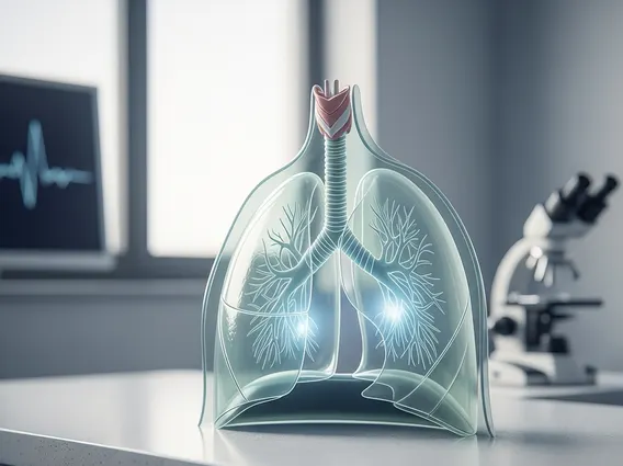
The Pleura refers to the serous membrane that envelops the lungs and lines the thoracic cavity. Each lung is enclosed by its own pleural sac, which is a double-layered membrane. This arrangement creates a sealed environment that is essential for the mechanics of breathing. The two layers, known as the visceral and parietal pleura, are separated by a potential space called the pleural cavity, which contains a thin film of pleural fluid.
This intricate structure ensures that the lungs can expand and contract smoothly within the chest cavity without friction. The integrity of the pleura and the balance of fluid within the pleural space are critical for normal respiratory function. Any disruption to this delicate system can lead to significant respiratory distress and various medical conditions.
Pleura Anatomy, Function, and Types
The pleura function and anatomy are intricately linked, enabling efficient respiration. Anatomically, the pleura is divided into two main layers:
- Visceral Pleura: This inner layer directly covers the surface of the lungs, extending into the fissures between the lobes. It is firmly attached to the lung tissue.
- Parietal Pleura: This outer layer lines the inside of the chest wall, the diaphragm, and the mediastinum (the space between the lungs). It is further subdivided based on the region it covers: costal (ribs), diaphragmatic (diaphragm), mediastinal (mediastinum), and cervical (neck).
Between these two layers lies the pleural cavity, a potential space containing a small amount of serous pleural fluid, typically 10-20 milliliters. The primary function of this fluid is to act as a lubricant, allowing the visceral and parietal layers to glide smoothly over each other during breathing, minimizing friction. Additionally, the fluid creates surface tension, causing the two pleural layers to adhere to each other, which is crucial for transmitting the negative pressure of the thoracic cavity to the lungs, facilitating their expansion.
The types of pleura are essentially these two distinct layers—visceral and parietal—each with specific anatomical relationships and roles in maintaining lung mechanics. Their combined action ensures the lungs remain inflated and can move freely within the chest.
Conditions and Diseases Affecting the Pleura
A range of pleura conditions and diseases can affect the integrity and function of these vital membranes, often leading to respiratory symptoms and pain. One common condition is pleurisy, also known as pleuritis, which is an inflammation of the pleura. This inflammation typically causes sharp chest pain that worsens with deep breaths, coughing, or sneezing. Pleurisy can result from viral infections, bacterial infections, autoimmune diseases, or even lung cancer.
Another significant condition is pleural effusion, characterized by an excessive accumulation of fluid in the pleural cavity. This can compress the lung, leading to shortness of breath and chest discomfort. Pleural effusions can be transudative (due to systemic conditions like heart failure or liver cirrhosis) or exudative (due to local inflammation or malignancy, such as pneumonia or cancer). According to the American Thoracic Society, approximately 1.5 million people in the United States are diagnosed with pleural effusion annually, highlighting its prevalence.
Pneumothorax, or a collapsed lung, occurs when air leaks into the pleural space, increasing pressure and causing the lung to partially or completely collapse. This can be spontaneous (without obvious cause), traumatic (due to injury), or iatrogenic (caused by medical procedures). Mesothelioma is a rare but aggressive cancer that originates in the pleura, often linked to asbestos exposure. These conditions underscore the importance of prompt diagnosis and treatment to preserve respiratory health and prevent severe complications.