
Pet Scan
A Positron Emission Tomography (PET) scan is a powerful diagnostic imaging tool used in medicine to visualize metabolic activity in the body. It plays a crucial role in detecting diseases, assessing their severity, and monitoring treatment effectiveness by revealing how organs and tissues are functioning at a cellular level.
Key Takeaways
- A PET scan utilizes a radioactive tracer to highlight metabolic activity, aiding in the detection of diseases at an early, cellular stage.
- It is an invaluable tool for diagnosing and staging various cancers, evaluating heart conditions, and investigating neurological disorders.
- The procedure involves injecting a small amount of tracer, which accumulates in areas of high metabolic activity, followed by imaging.
- Patients typically need to fast and adjust certain medications before a PET scan to ensure accurate results.
- PET scans provide unique functional insights into the body, complementing the structural information obtained from other imaging techniques like CT or MRI.
What is a PET scan (Positron Emission Tomography)?
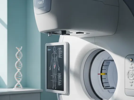
A PET scan, or Positron Emission Tomography scan, is an advanced nuclear medicine imaging technique that provides detailed information about the metabolic and biochemical function of tissues and organs. Unlike X-rays or CT scans, which primarily show anatomical structures, a PET scan reveals how well cells are working by detecting changes at a molecular level. This capability makes it particularly useful for identifying diseases in their earliest stages, often before structural changes become apparent on other imaging tests. When considering what is a PET scan, it’s essential to understand that it involves injecting a small amount of a radioactive tracer, typically a glucose analog like fluorodeoxyglucose (FDG), into the bloodstream. This tracer travels through the body and accumulates in cells that are highly metabolically active, such as cancer cells, inflamed tissues, or active brain regions.
The scanner then detects the energy emitted by the tracer, which is converted into three-dimensional images. These images highlight areas of abnormal metabolic activity, providing critical insights into disease presence, spread, and response to treatment. The information gathered from a PET scan can guide treatment decisions, help stage diseases, and monitor the effectiveness of therapies, offering a comprehensive view of a patient’s health status.
How PET Scans Work and Their Clinical Applications
The mechanism behind a PET scan involves the administration of a radiotracer, which is absorbed by metabolically active cells. Once absorbed, the tracer emits positrons, which collide with electrons in the body, producing gamma rays. These gamma rays are detected by the PET scanner, which then uses sophisticated computer software to reconstruct detailed images showing the distribution of the tracer throughout the body. Areas with higher tracer uptake indicate increased metabolic activity, which can be indicative of disease processes. This functional imaging capability is what makes PET scans so valuable in various medical fields.
The PET scan uses and benefits are extensive, particularly in oncology, cardiology, and neurology. In oncology, PET scans are widely used for:
- Detecting cancer and determining if it has spread (staging).
- Evaluating the effectiveness of cancer treatments by observing changes in tumor activity.
- Detecting cancer recurrence after treatment.
- Guiding biopsies to the most metabolically active parts of a tumor.
In cardiology, PET scans can assess blood flow to the heart muscle and identify areas of damaged heart tissue, helping to diagnose coronary artery disease and determine the viability of heart muscle after a heart attack. For neurological conditions, PET scans are instrumental in diagnosing Alzheimer’s disease by detecting amyloid plaques, localizing seizure foci in epilepsy, and evaluating movement disorders like Parkinson’s disease. The ability to visualize physiological processes rather than just anatomy provides a unique diagnostic advantage, often complementing information from other imaging modalities.
Preparing for Your PET Scan
Proper preparing for a PET scan is crucial to ensure the accuracy and effectiveness of the procedure. Patients will typically receive specific instructions from their healthcare provider, but general guidelines often include several key steps. Most commonly, patients are advised to fast for a certain period, usually 4-6 hours, before the scan. This is because the most common tracer, FDG, is a sugar molecule, and food intake can affect blood sugar levels, potentially altering tracer distribution and scan results, especially for diabetic patients.
Other important preparatory steps include:
- Medication Review: Discuss all current medications with your doctor, particularly those for diabetes, as adjustments might be necessary.
- Hydration: Drinking plenty of water is often encouraged, as good hydration can help the tracer distribute effectively and aid in its excretion.
- Physical Activity: Patients are usually asked to avoid strenuous exercise for 24 hours before the scan, as muscle activity can increase glucose uptake in muscles, potentially obscuring other areas of interest.
- Clothing: Wear comfortable, loose-fitting clothing without metal zippers or buttons, as metal can interfere with the imaging process.
- Relaxation: It’s important to remain still during the scan, so patients are often advised to try and relax before and during the procedure.
Following these instructions carefully helps ensure that the PET scan provides the clearest and most accurate diagnostic information possible, aiding in effective patient care and treatment planning.


