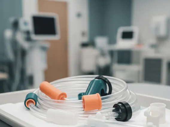
Percutaneous Endoscopic Tube
A Percutaneous Endoscopic Tube (PET) is a medical device inserted through the abdominal wall into the stomach or small intestine, primarily to provide nutritional support or medication when oral intake is not possible or safe. This procedure offers a vital solution for patients facing long-term feeding challenges.
Key Takeaways
- A Percutaneous Endoscopic Tube (PET) is a feeding tube placed through the skin into the gastrointestinal tract, often the stomach (PEG) or jejunum (PEJ).
- It is used for long-term nutritional support, medication delivery, or gastric decompression in patients unable to eat or swallow safely.
- The procedure involves endoscopic guidance, local anesthesia, and sedation, typically taking less than an hour.
- Recovery involves initial site care and gradual feeding, with most patients adapting well to the tube within a few days to weeks.
- Proper care of the tube site is crucial to prevent complications like infection.
What is a Percutaneous Endoscopic Tube?
A Percutaneous Endoscopic Tube (PET) refers to a type of feeding tube that is inserted through the skin (percutaneous) of the abdomen and directly into the stomach or small intestine, with the aid of an endoscope. This method allows for direct delivery of nutrition, fluids, and medications into the digestive system, bypassing the mouth and esophagus. The most common type is a Percutaneous Endoscopic Gastrostomy (PEG) tube, which goes into the stomach, but tubes can also be placed into the jejunum (Percutaneous Endoscopic Jejunostomy or PEJ).
PETs are crucial for individuals who are unable to swallow safely or adequately, such as those with severe dysphagia due to neurological conditions (e.g., stroke, Parkinson’s disease), head and neck cancers, or severe malnutrition. They provide a reliable and long-term solution for maintaining hydration and nutritional status, significantly improving quality of life for many patients. According to the American Society for Gastrointestinal Endoscopy, PEG tube placement is a common procedure, with hundreds of thousands performed annually worldwide, highlighting its importance in clinical practice.
Percutaneous Endoscopic Tube Procedure and Uses
The placement of a Percutaneous Endoscopic Tube is a minimally invasive procedure, typically performed under local anesthesia and conscious sedation. During the percutaneous endoscopic tube procedure explained, an endoscope is passed through the mouth, esophagus, and into the stomach to visualize the stomach wall. The physician then identifies an appropriate site on the abdominal wall, and a small incision is made. A needle is passed through the abdominal wall into the stomach, and a guidewire is threaded through the needle. The endoscope is used to grasp the guidewire and pull it out through the mouth. The feeding tube is then attached to the guidewire and pulled down through the mouth, esophagus, stomach, and out through the abdominal incision, where it is secured with an external bumper.
The percutaneous endoscopic tube uses and benefits are extensive, primarily centered around providing essential support when oral intake is compromised. Key applications include:
- Nutritional Support: Delivering liquid nutrition directly to the stomach or small intestine for patients with swallowing difficulties, chronic illnesses, or severe malnutrition.
- Medication Administration: Providing medications that cannot be taken orally or require precise dosing directly into the digestive tract.
- Gastric Decompression: In some cases, a PET can be used to drain excess air or fluid from the stomach, relieving nausea and vomiting.
The benefits include improved nutritional status, better hydration, enhanced medication efficacy, and a significant improvement in the overall quality of life by reducing the risk of aspiration pneumonia and ensuring adequate caloric intake.
Recovery and Care After Percutaneous Endoscopic Tube Placement
After Percutaneous Endoscopic Tube placement, patients typically experience a relatively quick initial recovery. Most individuals can resume light activities within a day or two, though it’s common to feel some discomfort or soreness at the insertion site. Pain management is usually achieved with over-the-counter pain relievers. The percutaneous endoscopic tube recovery time for full adaptation, including learning how to manage feeds and care for the site, can vary from a few days to several weeks, depending on the individual’s overall health and the underlying condition requiring the tube.
Proper care of the tube site is paramount to prevent complications such as infection or skin irritation. This involves daily cleaning of the skin around the tube with mild soap and water, ensuring the site remains dry, and regularly checking for signs of infection like redness, swelling, or discharge. Dressings may be required initially and should be changed as advised by healthcare providers. Patients and caregivers receive comprehensive training on how to administer feeds, flush the tube to prevent blockages, and recognize potential issues. Regular follow-up appointments are essential to monitor the tube’s function, assess nutritional status, and address any concerns.