
Pedicle Flap
A pedicle flap is a reconstructive surgical technique used to transfer tissue from one part of the body to another while maintaining its original blood supply. This method is crucial for repairing defects resulting from trauma, cancer removal, or congenital conditions, ensuring the transferred tissue remains viable.
Key Takeaways
- A pedicle flap involves moving tissue with its attached blood supply to reconstruct defects.
- This surgical method ensures the transferred tissue remains alive and healthy in its new location.
- Types of pedicle flaps vary based on their proximity to the defect, including local and regional options.
- The procedure involves careful dissection, transfer, and securement of the flap, followed by a recovery period focused on healing and monitoring.
- Pedicle flaps are vital in reconstructive surgery for restoring function and appearance after significant tissue loss.
What is a Pedicle Flap?
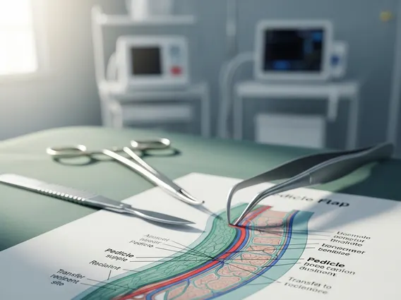
A Pedicle Flap is a fundamental technique in reconstructive surgery where a section of tissue, including skin, muscle, fat, or bone, is moved from a donor site to a recipient site. The defining characteristic of this procedure is that the tissue remains partially attached to its original location, preserving its blood supply through a vascular “pedicle.” This continuous blood flow is critical, as it nourishes the transferred tissue, preventing necrosis and ensuring its integration into the new site. The concept of what is a pedicle flap revolves around this principle of maintaining vascularity during tissue transfer, which is essential for successful reconstruction of complex defects, particularly in areas requiring robust, well-perfused tissue.
This surgical approach is widely utilized when direct closure of a wound is not possible, or when the defect requires a significant amount of tissue for coverage, volume restoration, or functional reconstruction. Unlike a free flap, which involves detaching the tissue completely and reattaching its blood vessels using microsurgery, a pedicle flap relies on the intact vascular connection, making it a simpler option in certain scenarios. The choice of flap depends on the size and location of the defect, the availability of donor tissue, and the patient’s overall health.
Types of Pedicle Flaps
There are several types of pedicle flaps, categorized primarily by their proximity to the defect and the nature of their vascular pedicle. Each type is chosen based on the specific reconstructive needs, the size and location of the defect, and the characteristics of the available donor tissue. Understanding these variations is crucial for effective surgical planning.
- Local Flaps: These flaps are harvested from tissue immediately adjacent to the defect. They are rotated, advanced, or transposed to cover the nearby wound. Examples include rotation flaps, advancement flaps, and transposition flaps. Local flaps typically offer excellent tissue match in terms of color and texture.
- Regional Flaps: These flaps involve tissue from a nearby, but not directly adjacent, anatomical region. They are often larger and can provide more bulk than local flaps. The pedicle, containing the blood vessels, is typically tunneled under intact skin to reach the recipient site. Common examples include the pectoralis major flap for head and neck reconstruction or the latissimus dorsi flap for breast reconstruction.
- Distant Pedicle Flaps: While less common today due to advancements in microsurgery for free flaps, distant pedicle flaps involve transferring tissue from a non-adjacent part of the body. Historically, these might involve creating a temporary attachment to a limb for several weeks until new blood supply formed, then detaching the original pedicle. More modern distant pedicle flaps rely on very long vascular pedicles or staged procedures.
The selection of a specific flap type is a critical decision made by the surgical team, considering factors such as the defect’s size, the need for specific tissue components (e.g., muscle, bone), and the potential impact on the donor site.
Pedicle Flap Procedure and Recovery
The pedicle flap procedure details involve several meticulous steps, beginning with careful assessment and planning. The surgeon first identifies a suitable donor site that can provide adequate tissue without compromising function or aesthetics. The design of the flap is critical, ensuring it contains a reliable vascular pedicle—a bundle of blood vessels that will sustain the tissue once it’s moved. This initial phase is vital for a successful pedicle flap surgery explanation.
During the surgery, the flap is carefully incised and elevated from the donor site, keeping its vascular pedicle intact. It is then rotated, advanced, or tunneled to the recipient site, where the defect needs to be covered. Once positioned, the flap is meticulously sutured into place, ensuring good contact and tension-free closure. The donor site is then closed, either directly or with a skin graft, depending on its size and location. The entire process demands precision to maintain blood flow and achieve optimal functional and aesthetic outcomes.
Recovery from a pedicle flap procedure typically involves a hospital stay, during which the flap’s viability is closely monitored. Surgeons and nurses regularly check the flap’s color, temperature, and capillary refill to ensure adequate blood supply. Pain management, wound care, and infection prevention are also critical components of the post-operative period. Patients are usually advised to avoid strenuous activities and protect the surgical sites for several weeks. Full healing can take several months, and rehabilitation, such as physical therapy, may be necessary to restore function, especially if muscle or bone was transferred. Long-term follow-up is essential to monitor the flap’s integration and address any potential complications or aesthetic concerns.


