Optical Spectroscopy
Optical Spectroscopy is a powerful analytical technique that utilizes the interaction of light with matter to gather information about its composition, structure, and properties. In clinical and medical contexts, it serves as a non-invasive tool for diagnosing diseases, monitoring physiological changes, and guiding therapeutic interventions.
Key Takeaways
- Optical Spectroscopy analyzes how light interacts with biological tissues to provide insights into their molecular and structural characteristics.
- Its fundamental principles involve measuring light absorption, scattering, and emission across various wavelengths.
- This technique is non-invasive, offering a significant advantage for patient comfort and repeated measurements.
- Key applications include early cancer detection, monitoring disease progression, and guiding surgical procedures.
- It holds promise for advancing personalized medicine and improving diagnostic accuracy in numerous medical fields.
What is Optical Spectroscopy?
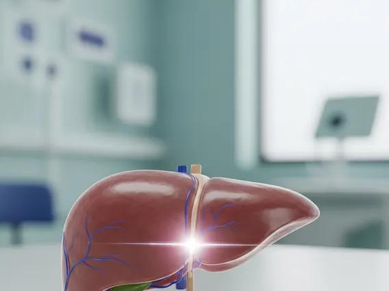
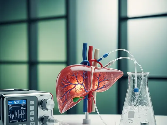
Optical Spectroscopy refers to a broad range of techniques that study the interaction between light (electromagnetic radiation) and biological samples. In the medical field, this interaction provides crucial information about tissue health, cellular metabolism, and molecular composition without the need for invasive biopsies. By analyzing how light is absorbed, scattered, or emitted by tissues, clinicians can identify subtle changes indicative of disease, such as alterations in blood flow, oxygenation levels, or the presence of specific biomarkers.
This non-destructive approach allows for real-time assessment, making it particularly valuable for applications requiring immediate feedback, like during surgical procedures or continuous patient monitoring. The technique leverages different parts of the electromagnetic spectrum, including ultraviolet (UV), visible, and near-infrared (NIR) light, each offering unique insights into biological processes at varying depths and resolutions.
Principles and Mechanisms of Optical Spectroscopy
The fundamental principles of optical spectroscopy are rooted in how photons interact with the molecules and structures within biological tissue. When light illuminates a sample, it can undergo several processes: absorption, scattering, and fluorescence. Each of these interactions provides distinct information. Absorption occurs when molecules absorb specific wavelengths of light, revealing their chemical composition. For instance, hemoglobin absorbs light differently depending on its oxygenation state, which is crucial for assessing tissue oxygenation.
Understanding how optical spectroscopy works involves recognizing these interactions. Scattering, on the other hand, occurs when light changes direction due to encountering cellular structures, organelles, or collagen fibers. The pattern and intensity of scattered light can provide information about tissue morphology and microstructure, which often changes during disease progression, such as in tumor growth. Fluorescence spectroscopy involves exciting molecules with light at one wavelength and then detecting the light they emit at a longer wavelength. Endogenous fluorophores, like NADH and flavins, are involved in cellular metabolism, and their fluorescence can indicate metabolic state changes associated with various pathologies.
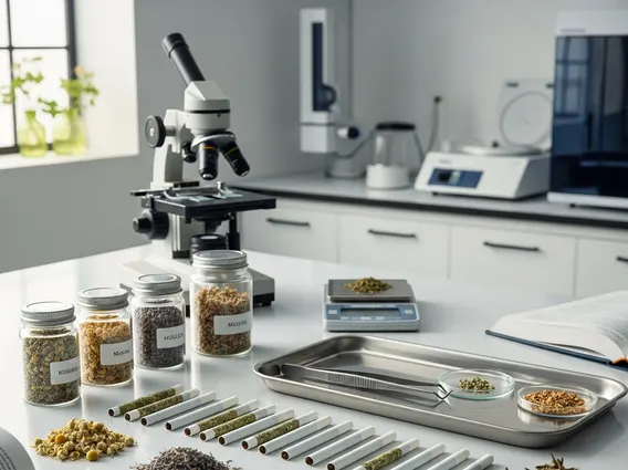
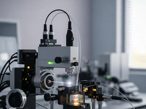
The instrumentation typically includes a light source (e.g., lasers, LEDs), optics to deliver light to the tissue and collect the interacting light, and a detector (e.g., spectrometer, camera) to measure the spectral properties. Sophisticated algorithms then process this spectral data to extract meaningful biological and clinical parameters.
Key Applications of Optical Spectroscopy
The applications of optical spectroscopy are diverse and rapidly expanding within medical diagnostics and therapy. One of its most significant roles is in early cancer detection, where it can differentiate between healthy and cancerous tissues based on their distinct optical signatures. For example, studies have have shown its potential in detecting early-stage cancers in the skin, cervix, and gastrointestinal tract, often providing results in real-time without the need for tissue removal.
Beyond diagnostics, optical spectroscopy is also employed for:
- Surgical Guidance: Helping surgeons delineate tumor margins more precisely during operations, ensuring complete removal while preserving healthy tissue.
- Treatment Monitoring: Assessing the effectiveness of therapies, such as photodynamic therapy or chemotherapy, by observing changes in tissue properties over time.
- Non-invasive Monitoring: Continuous monitoring of physiological parameters like blood glucose levels, tissue oxygenation, and cerebral hemodynamics, particularly in critical care settings.
- Pathology and Histology: Providing rapid, label-free assessment of tissue biopsies, potentially reducing the time and resources required for traditional histopathological analysis.
The ability of optical spectroscopy to provide detailed molecular and structural information non-invasively makes it an invaluable tool for advancing personalized medicine, enabling earlier diagnosis, and facilitating more targeted and effective treatments across a wide range of medical conditions.