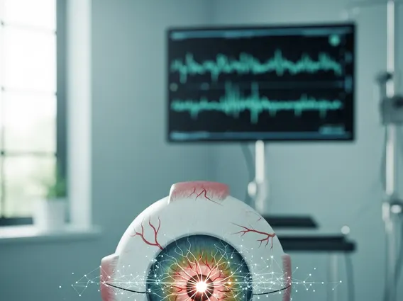
Optic Neuritis
Optic Neuritis is an inflammatory condition affecting the optic nerve, which transmits visual information from the eye to the brain. This inflammation can lead to sudden, temporary vision loss and pain, often signaling an underlying neurological condition.
Key Takeaways
- Optic Neuritis is an inflammation of the optic nerve, crucial for vision.
- It commonly causes sudden vision loss, pain with eye movement, and altered color perception.
- Often associated with demyelinating diseases like multiple sclerosis (MS).
- Diagnosis involves neurological and ophthalmological exams, including MRI.
- Treatment typically involves corticosteroids to reduce inflammation and speed recovery.
What is Optic Neuritis: Causes and Risk Factors
Optic Neuritis refers to inflammation that damages the optic nerve, a bundle of nerve fibers responsible for transmitting visual signals from the retina to the brain. This damage can disrupt the flow of visual information, leading to various degrees of vision impairment. The condition is often unilateral, affecting one eye, but can occasionally occur in both eyes.
The precise mechanisms behind optic neuritis symptoms causes are complex, but it is frequently linked to demyelination, a process where the protective myelin sheath around nerve fibers is damaged. The most common cause is multiple sclerosis (MS), an autoimmune disease that attacks the central nervous system. In fact, optic neuritis is often the first symptom of MS for many individuals, with approximately 50% of people with MS experiencing an episode of optic neuritis at some point. Other potential causes and risk factors include:
- Autoimmune Diseases: Beyond MS, conditions like neuromyelitis optica (NMO) and myelin oligodendrocyte glycoprotein antibody-associated disease (MOGAD) can cause optic neuritis.
- Infections: Viral infections such as measles, mumps, rubella, herpes, and Lyme disease, as well as bacterial infections like syphilis, can trigger optic neuritis.
- Other Inflammatory Conditions: Sarcoidosis, lupus, and Behçet’s disease are systemic inflammatory conditions that may affect the optic nerve.
- Medications: Certain drugs, including some antibiotics and antituberculosis medications, have been associated with optic neuritis as a rare side effect.
While optic neuritis can affect anyone, it is most common in adults between the ages of 20 and 45 and is more prevalent in women than men. A genetic predisposition may also play a role, as individuals with a family history of MS may have an increased risk.
Optic Neuritis Symptoms and Diagnosis
The symptoms of Optic Neuritis typically develop rapidly over hours or days. The most common symptom is sudden vision loss, which can range from a blurred spot to complete blindness in the affected eye. This vision loss often worsens over several days before improving. Another hallmark symptom is pain, especially with eye movement, which can precede or accompany the vision changes. Patients may also experience dyschromatopsia, a diminished ability to perceive colors, particularly red saturation, making colors appear faded or washed out.
How is Optic Neuritis Diagnosed?
Diagnosing Optic Neuritis involves a comprehensive evaluation by an ophthalmologist and often a neurologist. The diagnostic process typically includes a detailed medical history and a thorough eye examination. Key diagnostic steps include:
- Visual Acuity Test: Measures the sharpness of vision.
- Color Vision Test: Assesses the ability to distinguish colors, often revealing a deficit in red-green perception.
- Pupil Light Reflex Test: Checks for a relative afferent pupillary defect (RAPD), where the affected pupil constricts less when light is shone into it compared to the unaffected eye.
- Ophthalmoscopy: Examination of the back of the eye to check for swelling of the optic disc (papillitis), though in many cases, the optic nerve head may appear normal (retrobulbar optic neuritis).
- Magnetic Resonance Imaging (MRI): An MRI of the brain and orbits is crucial. It can detect inflammation of the optic nerve and identify demyelinating lesions in the brain, which are indicative of multiple sclerosis. According to the National Institute of Neurological Disorders and Stroke (NINDS), about 50% of people who experience optic neuritis develop MS within 15 years, especially if MRI shows brain lesions.
- Blood Tests: May be performed to rule out other causes, such as infections or specific autoimmune conditions like NMO or MOGAD.
The combination of clinical symptoms and objective findings from these tests helps confirm the diagnosis and guide further management.
Optic Neuritis Treatment Options
The primary goal of optic neuritis treatment options is to speed recovery of vision and reduce inflammation. While vision often recovers spontaneously over several weeks or months, treatment can accelerate this process and potentially reduce the risk of future episodes, especially in individuals at high risk for MS. The most common treatment involves corticosteroids.
High-dose intravenous corticosteroids, such as methylprednisolone, are typically administered for a few days, followed by oral corticosteroids that are gradually tapered. This treatment helps to reduce inflammation and swelling of the optic nerve. While corticosteroids can hasten visual recovery, studies have shown they do not significantly affect the final visual outcome or prevent future episodes of optic neuritis in the long term, though they may delay the onset of MS in some cases. For individuals with severe vision loss or those who do not respond to corticosteroids, plasma exchange (plasmapheresis) may be considered.
For patients diagnosed with or at high risk for multiple sclerosis, disease-modifying therapies (DMTs) may be initiated. These treatments aim to reduce the frequency and severity of MS relapses, including optic neuritis, and slow the progression of the disease. The choice of DMT depends on various factors, including the specific diagnosis (e.g., MS, NMO, MOGAD), disease activity, and individual patient characteristics. Regular follow-up with an ophthalmologist and neurologist is essential to monitor vision, manage symptoms, and adjust treatment as needed.