
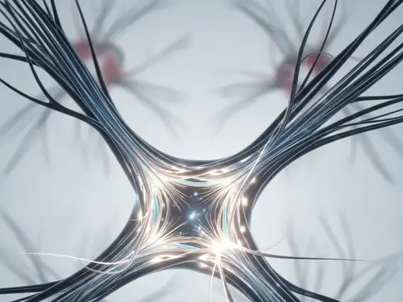
Optic Chiasm
The optic chiasm is a crucial structure in the human brain, playing a vital role in vision by facilitating the crossing of nerve fibers from the eyes. Understanding its anatomy and function is essential for comprehending how visual information is processed.
Key Takeaways
- The optic chiasm is an X-shaped structure where optic nerves from both eyes partially cross.
- It is located at the base of the brain, anterior to the pituitary gland.
- Its primary function is to enable visual information from the nasal (inner) half of each retina to cross to the opposite side of the brain.
- This crossing ensures that each side of the brain receives visual input from the contralateral visual field.
- Various conditions, including tumors and inflammatory diseases, can affect the optic chiasm, leading to characteristic visual field defects.
What is the Optic Chiasm: Anatomy and Function
The optic chiasm is a unique, X-shaped anatomical structure located at the base of the brain, just anterior to the pituitary gland and inferior to the hypothalamus. It is a critical component of the visual pathway where nerve fibers from the optic nerves partially decussate, or cross over, before continuing as the optic tracts.
The primary optic chiasm anatomy and function involve the organized crossing of nerve fibers. Specifically, fibers originating from the nasal (inner) half of each retina cross to the opposite side of the brain, while fibers from the temporal (outer) half of each retina remain on the same side. This arrangement is fundamental to binocular vision and depth perception. The optic chiasm location and purpose are integral to ensuring that visual information from the right half of the visual field (seen by the nasal retina of the right eye and the temporal retina of the left eye) is processed by the left cerebral hemisphere, and vice versa for the left visual field.
Key aspects of the optic chiasm’s function include:
- Partial Decussation: Only a portion of the optic nerve fibers cross, specifically those from the nasal retinas.
- Visual Field Integration: It ensures that each cerebral hemisphere receives a complete representation of the contralateral visual field.
- Binocular Vision: This crossing is essential for combining images from both eyes into a single, three-dimensional perception.
Conditions Affecting the Optic Chiasm
A variety of conditions affecting optic chiasm can lead to significant visual disturbances, often characterized by specific patterns of visual field loss. Due to its strategic location near the pituitary gland and hypothalamus, the optic chiasm is vulnerable to compression or damage from adjacent structures. The most common cause of chiasmal compression is a pituitary adenoma, a benign tumor of the pituitary gland, which can grow upwards and press directly on the chiasm.
Other conditions that can impact the optic chiasm include:
- Craniopharyngiomas: These are rare, benign brain tumors that develop near the pituitary stalk and can compress the chiasm.
- Meningiomas: Tumors arising from the meninges (membranes surrounding the brain and spinal cord) can also grow in the region of the optic chiasm.
- Aneurysms: Enlargements of blood vessels in the Circle of Willis, particularly the anterior communicating artery, can press on the chiasm.
- Inflammatory and Demyelinating Diseases: Conditions like multiple sclerosis or sarcoidosis can cause inflammation or demyelination of the optic chiasm, disrupting nerve signal transmission.
Damage to the optic chiasm typically results in bitemporal hemianopsia, a characteristic visual field defect where vision is lost in the outer halves of both visual fields. Diagnosis usually involves a combination of visual field testing, neurological examination, and imaging studies such as Magnetic Resonance Imaging (MRI) of the brain. Treatment depends on the underlying cause and may include surgical removal of tumors, radiation therapy, or medical management for inflammatory conditions.


