Ophthalmoscope
An ophthalmoscope is a vital diagnostic instrument used by healthcare professionals to examine the interior structures of the eye, particularly the retina, optic disc, macula, and blood vessels. This examination, known as ophthalmoscopy or funduscopy, is crucial for detecting various ocular and systemic diseases.
Key Takeaways
- An ophthalmoscope is a medical device used to visualize the internal structures of the eye, especially the retina.
- It helps diagnose conditions like glaucoma, diabetic retinopathy, and macular degeneration.
- The device works by emitting a light beam into the eye, allowing the examiner to view the reflected light through a system of lenses.
- There are primarily two types: direct ophthalmoscopes for magnified, detailed views and indirect ophthalmoscopes for a wider field of view.
- The examination is a non-invasive procedure, often involving pupil dilation, to assess eye health comprehensively.
What is an Ophthalmoscope and Its Clinical Applications?
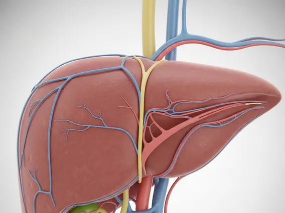
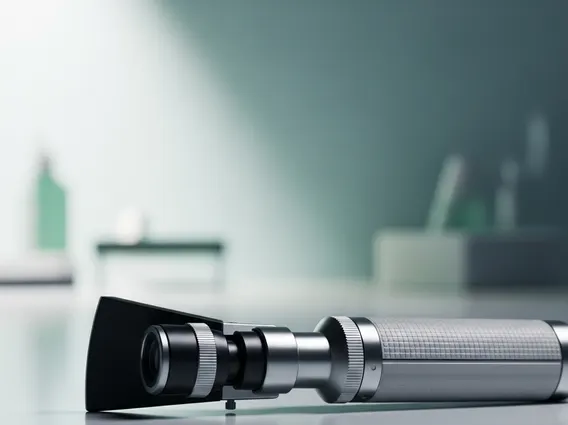
An Ophthalmoscope is a handheld or head-mounted medical device designed to illuminate and magnify the internal structures of the eye. Its primary purpose is to allow clinicians to inspect the fundus, which includes the retina, optic disc, macula, and choroid. This instrument is indispensable in ophthalmology and general medicine for assessing ocular health and detecting signs of various diseases.
The clinical applications of an ophthalmoscope are extensive. It is used for identifying conditions such as diabetic retinopathy, where high blood sugar damages retinal blood vessels; glaucoma, characterized by optic nerve damage often linked to increased intraocular pressure; and macular degeneration, which affects central vision. Furthermore, it can reveal signs of systemic diseases like hypertension (hypertensive retinopathy), brain tumors (papilledema), and even certain infectious diseases. Early detection through regular ophthalmoscopic examinations can significantly impact treatment outcomes and preserve vision.
How an Ophthalmoscope Works and Its Different Types
An ophthalmoscope works by projecting a beam of light into the patient’s eye, which then reflects off the retina. The examiner looks through a small aperture in the device, utilizing a series of lenses and mirrors to focus on the illuminated structures. The instrument typically includes a selection of lenses that can be rotated to correct for refractive errors in both the patient’s and examiner’s eyes, allowing for a clear, magnified view of the fundus.
There are two main Types of ophthalmoscopes and their uses:
- Direct Ophthalmoscope: This is the most common type, providing an upright, magnified image (approximately 15x) of a small area of the retina. It is ideal for detailed examination of specific retinal lesions, the optic disc, and the macula. The examiner holds the device close to the patient’s eye.
- Indirect Ophthalmoscope: This type, often head-mounted, uses a separate condensing lens held in front of the patient’s eye. It provides an inverted, less magnified (2-5x) but much wider field of view of the retina. Indirect ophthalmoscopy is particularly useful for assessing the peripheral retina and for patients with small pupils or media opacities.
Each type offers distinct advantages, and clinicians often use both depending on the specific diagnostic needs. For instance, an indirect ophthalmoscope might be used for an initial comprehensive survey, followed by a direct ophthalmoscope for closer inspection of suspicious areas.
The Ophthalmoscope Exam Procedure Explained
The Ophthalmoscope exam procedure explained typically begins with the patient seated comfortably in a darkened room. Often, eye drops are administered to dilate the pupils, which allows for a wider and clearer view of the fundus. This dilation usually takes about 15-30 minutes to take effect and can cause temporary light sensitivity and blurred vision.
During a direct ophthalmoscopy, the examiner sits facing the patient, holding the ophthalmoscope close to their own eye and about 1-2 inches from the patient’s eye. The examiner directs the light beam into the patient’s pupil, adjusting the focus wheel to bring the retinal structures into sharp view. They systematically examine different areas of the retina, noting the color and clarity of the optic disc, the condition of blood vessels, and the presence of any abnormalities in the macula or peripheral retina. For indirect ophthalmoscopy, the examiner wears a head-mounted device and holds a condensing lens in front of the patient’s eye, viewing the inverted image through the device. The entire procedure is generally painless, though the bright light can be momentarily uncomfortable. Regular ophthalmoscopic examinations are a cornerstone of preventative eye care, recommended by organizations like the American Academy of Ophthalmology, especially for individuals at risk of eye diseases.