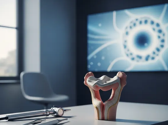
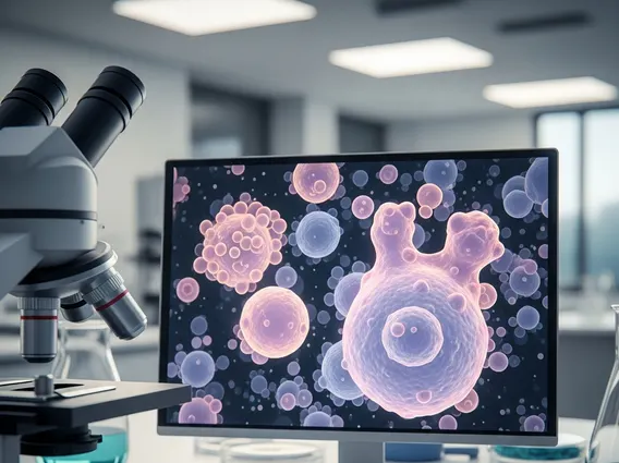
Nuclear Medicine
Nuclear Medicine is a specialized medical field that uses small, safe amounts of radioactive materials, known as radiopharmaceuticals or radiotracers, to diagnose and treat a wide range of diseases. This unique approach provides insights into organ function and structure, offering critical information often unavailable through other imaging techniques.
Key Takeaways
- Nuclear Medicine utilizes radiopharmaceuticals to visualize organ function and structure.
- It helps diagnose and treat various conditions across cardiology, oncology, endocrinology, and neurology.
- Procedures are generally non-invasive and provide functional information about the body.
- Common imaging techniques include PET (Positron Emission Tomography) and SPECT (Single-Photon Emission Computed Tomography) scans.
- The field focuses on physiological processes, making it distinct from anatomical imaging methods like X-rays or CT scans.
What is Nuclear Medicine?
Nuclear Medicine is a distinct medical specialty that employs radioactive substances to assess bodily functions and to diagnose and treat disease. Unlike traditional imaging methods that primarily show anatomy, nuclear medicine focuses on visualizing physiological processes at a cellular and molecular level. This allows clinicians to detect diseases in their earliest stages, often before structural changes become apparent on other imaging tests.
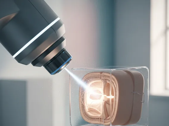
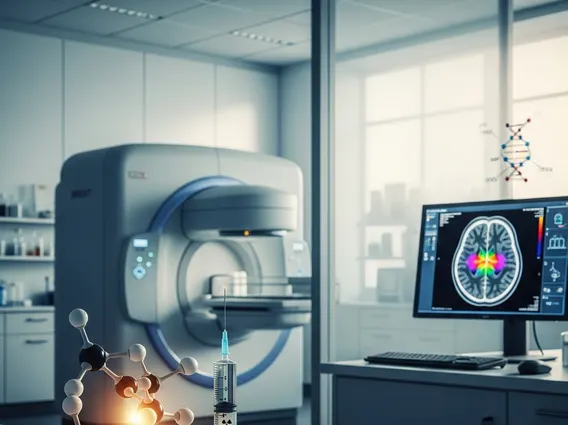
The core principle involves administering a radiotracer, which is a pharmaceutical combined with a small amount of a radioactive isotope. This radiotracer is designed to target specific organs, tissues, or cells within the body. Once administered, it emits gamma rays, which are then detected by specialized cameras to create detailed images or measure organ function.
Mechanism, Uses, and Common Procedures
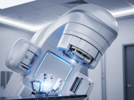
Understanding how nuclear medicine works involves the journey of these radiotracers. After administration, typically through injection, ingestion, or inhalation, the radiotracer travels through the body and accumulates in the target area. The specific radiotracer chosen depends on the organ or function being studied. For example, some tracers are absorbed by metabolically active cells, while others bind to specific receptors or track blood flow. Specialized cameras, such as SPECT (Single-Photon Emission Computed Tomography) or PET (Positron Emission Tomography) scanners, detect the emitted gamma rays. These signals are then processed by computers to generate detailed 2D or 3D images, providing functional information about the body’s systems.
The uses of nuclear medicine are extensive, spanning numerous medical disciplines for both diagnostic and therapeutic purposes. Diagnostically, it is invaluable for:
- Cardiology: Assessing blood flow to the heart muscle, detecting coronary artery disease, and evaluating heart function.
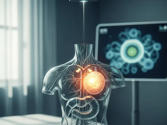
- Oncology: Identifying cancerous tumors, staging cancer, determining the spread of cancer (metastasis), and monitoring response to treatment.
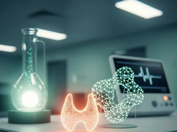
- Endocrinology: Evaluating thyroid function, locating parathyroid adenomas, and assessing adrenal gland disorders.
- Neurology: Diagnosing conditions like Alzheimer’s disease, Parkinson’s disease, and seizure disorders, as well as evaluating brain blood flow.
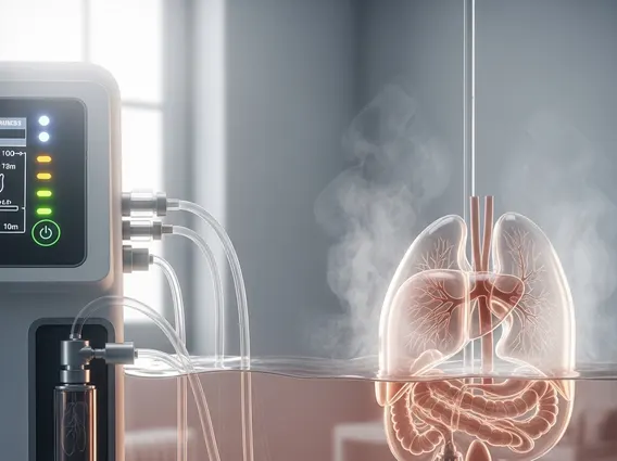
- Bone: Detecting fractures, infections, and tumors that may not be visible on standard X-rays.
Therapeutically, nuclear medicine uses higher doses of radiopharmaceuticals to deliver targeted radiation directly to diseased cells, such as in the treatment of thyroid cancer or certain neuroendocrine tumors.
Several nuclear medicine procedures explained are routinely performed to aid in diagnosis and treatment planning. The most common imaging modalities include PET and SPECT scans. PET scans often use a glucose-based radiotracer (FDG) to highlight areas of high metabolic activity, which is characteristic of many cancers and certain neurological conditions. SPECT scans provide three-dimensional images and are frequently used for cardiac stress tests, bone scans, and brain imaging. Other procedures include thyroid scans to assess gland function and detect nodules, lung scans to evaluate pulmonary embolism, and kidney scans to assess renal function and blood flow. These procedures are generally non-invasive and provide unique insights into the body’s functional status, making nuclear medicine a crucial tool in modern healthcare.