
Needle Wire Localization
Needle wire localization is a crucial medical procedure used to precisely mark the location of abnormal tissue, particularly non-palpable lesions, within the body before surgical removal. This technique ensures accurate targeting for biopsies or excisions, minimizing the removal of healthy surrounding tissue.
Key Takeaways
- Needle wire localization accurately marks non-palpable lesions for surgical removal.
- It is commonly employed in breast cancer diagnosis and treatment, guiding surgeons to abnormalities.
- The procedure involves imaging guidance (mammography, ultrasound) to place a thin wire into the target tissue.
- Benefits include precise lesion removal and preservation of healthy tissue.
- Potential risks are generally minor, such as discomfort or wire displacement.
What is Needle Wire Localization: Purpose and Procedure Overview
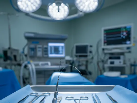
Needle Wire Localization refers to a medical procedure designed to pinpoint the exact location of an abnormality, such as a tumor or suspicious lesion, that cannot be felt during a physical examination. This technique is predominantly used in oncology, especially for breast lesions, to guide surgeons to the precise area requiring removal. The primary purpose is to ensure that the entire abnormal tissue is excised while preserving as much healthy surrounding tissue as possible.
The procedure typically involves using an imaging modality, such as mammography, ultrasound, or MRI, to visualize the target lesion. Once the lesion is accurately identified, a thin, flexible wire with a small hook or barb at its tip is carefully inserted through a needle and positioned directly into or adjacent to the abnormality. The needle is then removed, leaving the wire in place. This wire acts as a roadmap for the surgeon, guiding them directly to the target area during the subsequent surgical procedure. Understanding needle wire localization technique is essential for both medical professionals and patients, as it directly impacts the success and precision of lesion removal.
How Needle Wire Localization Works for Breast Biopsy
Needle wire localization plays a vital role in breast biopsy and surgical excision, particularly for non-palpable breast lesions detected through screening mammograms or other imaging studies. The process begins with the patient positioned appropriately for the chosen imaging guidance. For instance, if mammography is used, the breast is compressed, and images are taken to guide the needle. If ultrasound is preferred, gel is applied to the skin, and the transducer provides real-time visualization.
Under continuous imaging guidance, a local anesthetic is administered to numb the skin and deeper tissues. A thin needle, containing the localization wire, is then advanced towards the target lesion. The physician carefully monitors the needle’s path to ensure accurate placement. Once the tip of the needle is confirmed to be within or immediately adjacent to the lesion, the localization wire is deployed, and the needle is withdrawn, leaving the wire securely anchored. The external portion of the wire is taped to the skin and covered, ready for the surgeon to follow during the biopsy or lumpectomy. This method significantly enhances the surgeon’s ability to locate and remove the specific area of concern with high precision.
Benefits and Risks of Needle Wire Localization
Needle wire localization offers significant advantages, particularly in the context of breast cancer diagnosis and treatment. The primary benefit is the ability to precisely locate and remove non-palpable lesions, which are often early-stage cancers or suspicious abnormalities that cannot be felt by hand. This precision helps in achieving clear surgical margins, meaning that all cancerous cells are removed, reducing the need for repeat surgeries. Additionally, it minimizes the removal of healthy breast tissue, leading to better cosmetic outcomes and preserving breast volume. The procedure also allows for targeted biopsies, ensuring that the tissue sample obtained is representative of the abnormality.
While generally safe, needle wire localization benefits and risks should be carefully considered. Potential risks are typically minor and temporary. These may include:
- Discomfort or Pain: Mild pain or bruising at the insertion site is common.
- Bleeding: Minor bleeding can occur, usually resolving on its own.
- Infection: Though rare, there is a small risk of infection at the wire insertion site.
- Wire Displacement: In infrequent cases, the wire may shift slightly from its intended position, potentially requiring repositioning.
- Vasovagal Reaction: Some patients may experience lightheadedness or fainting during the procedure.
Patients are usually monitored for a short period after the procedure to ensure stability before proceeding to surgery. The overall benefits of accurate lesion localization for diagnosis and treatment generally outweigh these potential, usually manageable, risks.


