
Needle Localization
Needle Localization is a crucial medical procedure used to precisely mark the location of non-palpable lesions or abnormalities within the body, most commonly in the breast, before surgical removal or biopsy. This technique ensures accurate targeting, minimizing the removal of healthy tissue and improving diagnostic and therapeutic outcomes.
Key Takeaways
- Needle Localization is a pre-surgical or pre-biopsy procedure to mark non-palpable lesions.
- It uses imaging guidance (mammography, ultrasound, MRI) to place a thin wire or marker precisely.
- The technique is vital for ensuring accurate removal of abnormal tissue while preserving healthy surrounding tissue.
- It is most frequently employed in breast biopsies for lesions not detectable by touch.
- The procedure enhances the success rate of biopsies and surgical excisions by guiding the surgeon directly to the target area.
What is Needle Localization?
Needle Localization refers to a medical procedure designed to accurately pinpoint the exact location of an abnormality within the body that cannot be felt or seen during a physical examination. This technique is essential for guiding surgeons to the precise area requiring biopsy or surgical removal, particularly for small or deeply embedded lesions. The primary goal is to ensure that the entire abnormal tissue is removed while minimizing damage to surrounding healthy tissue. An effective understanding needle localization technique is critical for radiologists and surgeons alike, as it directly impacts the success and precision of subsequent medical interventions.
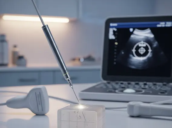
This procedure typically involves the use of imaging guidance, such as mammography, ultrasound, or magnetic resonance imaging (MRI), to visualize the lesion. Once the abnormality is located, a thin, flexible wire or a small marker is inserted through a needle and positioned within or adjacent to the target area. The external portion of the wire is then secured to the skin, providing a clear path for the surgeon. This precise marking is invaluable for lesions that are too small or too deep to be located by touch, ensuring that the diagnostic or therapeutic procedure is performed with the highest possible accuracy.
The Needle Localization Procedure: How It Works
The Needle Localization procedure involves several careful steps to ensure accurate placement of the guiding wire. It begins with the patient positioned comfortably, and the area to be localized is prepared with antiseptic. Imaging guidance is then used to visualize the lesion in real-time. For breast lesions, mammography or ultrasound are common, while MRI may be used for more complex cases or when other imaging modalities are insufficient. The radiologist carefully guides a thin needle towards the target abnormality.
Once the needle tip is confirmed to be within or immediately adjacent to the lesion, a specialized localization wire, often with a barb or hook at its tip, is threaded through the needle. The needle is then carefully withdrawn, leaving the wire in place. The wire’s tip anchors itself within the tissue, preventing displacement. The external part of the wire is secured to the skin with tape and covered with a dressing. This wire serves as a roadmap for the surgeon, leading them directly to the lesion during the subsequent biopsy or surgical excision. Post-procedure imaging may be performed to confirm the wire’s position before the patient proceeds to surgery.
Key steps in the procedure include:
- Imaging Guidance: Utilizing mammography, ultrasound, or MRI to precisely locate the lesion.
- Local Anesthesia: Administering a local anesthetic to numb the skin and deeper tissues at the needle insertion site.
- Needle Insertion: Guiding a thin needle to the target lesion under continuous imaging.
- Wire Placement: Advancing a localization wire through the needle, positioning its tip within or next to the abnormality.
- Needle Removal: Carefully withdrawing the needle, leaving the wire securely anchored in the tissue.
- Securing the Wire: Taping the external portion of the wire to the skin and applying a sterile dressing.
- Confirmation Imaging: Performing additional imaging to verify the wire’s final position relative to the lesion.
Needle Localization for Breast Biopsy
Needle Localization for breast biopsy is a widely adopted technique, particularly for lesions that are non-palpable, meaning they cannot be felt during a physical breast exam. These often include microcalcifications, architectural distortions, or small masses detected on mammograms, ultrasounds, or MRIs. The precision offered by needle localization is paramount in breast cancer diagnosis, as it allows surgeons to accurately remove the suspicious area for pathological examination while preserving as much healthy breast tissue as possible.
According to the American Cancer Society, breast cancer is the second most common cancer among women in the United States, and early detection through screening often identifies abnormalities that require further investigation, such as a biopsy. Many of these abnormalities are non-palpable, making needle localization an indispensable tool. By guiding the surgeon directly to the area of concern, the procedure significantly increases the likelihood of obtaining an adequate tissue sample for diagnosis, thereby improving patient outcomes and reducing the need for repeat procedures. This targeted approach is crucial for both diagnostic biopsies and therapeutic excisions, ensuring that the entire suspicious area is removed for definitive assessment.


