Myeloid Sarcoma
Myeloid sarcoma is a rare extramedullary tumor composed of immature myeloid cells, which are precursors to white blood cells. This condition is often associated with acute myeloid leukemia (AML) or other myeloproliferative neoplasms, but can also occur independently.
Key Takeaways
- Myeloid Sarcoma is a rare tumor of immature myeloid cells, often linked to acute myeloid leukemia (AML).
- It can manifest in various body sites, including skin, lymph nodes, bones, and central nervous system.
- Diagnosis relies on biopsy and immunohistochemistry, confirming the presence of myeloid blast cells.
- Treatment typically mirrors that of AML, involving intensive chemotherapy, with radiation and surgery for localized disease.
- Prognosis varies depending on its association with underlying hematologic conditions and the extent of the disease.
What is Myeloid Sarcoma?
Myeloid Sarcoma refers to a rare solid tumor composed of immature myeloid cells, also known as myeloblasts or granulocytic sarcoma. These tumors can appear in various parts of the body outside the bone marrow, blood, or lymph nodes, making their presentation highly variable. While it can occur de novo, it is most frequently observed in patients with or developing acute myeloid leukemia (AML), myelodysplastic syndromes (MDS), or myeloproliferative neoplasms (MPN). Its incidence is relatively low, estimated to occur in approximately 2-5% of patients with AML, according to studies published in medical journals like Blood Cancer Journal.
The presence of Myeloid Sarcoma often indicates a more aggressive disease course in patients with underlying hematologic conditions. The tumor cells are identical to those found in the bone marrow of individuals with AML, but they form a distinct mass in extramedullary sites. Common locations for these tumors include the skin (presenting as chloroma), lymph nodes, bones, soft tissues, and the central nervous system, though virtually any organ can be affected.
Clinical Presentation, Causes, and Diagnosis of Myeloid Sarcoma
The clinical presentation of myeloid sarcoma symptoms and signs is highly diverse, depending on the tumor’s location. For instance, skin involvement may present as firm, greenish (chloroma) or grayish-red nodules. Orbital involvement can lead to proptosis, while spinal cord compression may cause neurological deficits. Other common sites include lymph nodes, gastrointestinal tract, and reproductive organs, leading to symptoms specific to those areas, such as abdominal pain or organ dysfunction.
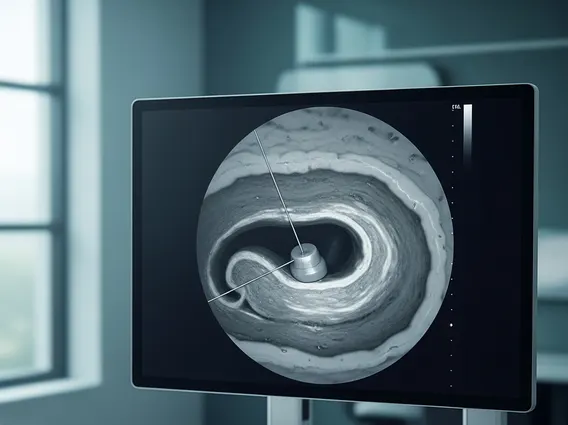
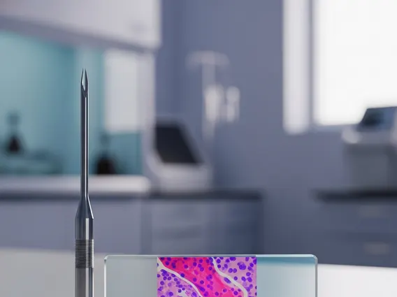
The exact myeloid sarcoma causes and diagnosis are complex. While the precise trigger for myeloid cells to form extramedullary tumors is not fully understood, it is strongly linked to underlying myeloid malignancies. Genetic mutations commonly found in AML, such as FLT3, NPM1, and CEBPA, are also often present in myeloid sarcoma cells. Diagnosis primarily relies on biopsy of the suspected mass, followed by histopathological examination and immunohistochemistry. These tests confirm the presence of myeloid blast cells and differentiate them from other types of tumors. Imaging techniques like CT, MRI, and PET scans are crucial for identifying the extent and location of the tumors.
Diagnostic methods for Myeloid Sarcoma typically involve a combination of approaches:
| Diagnostic Method | Purpose | Key Findings |
|---|---|---|
| Biopsy and Histopathology | To obtain tissue for microscopic examination. | Presence of immature myeloid cells (myeloblasts). |
| Immunohistochemistry | To identify specific markers on tumor cells. | Positive for myeloid markers (e.g., CD33, CD117, MPO). |
| Cytogenetics and Molecular Testing | To detect chromosomal abnormalities and gene mutations. | Common AML-associated mutations (e.g., FLT3, NPM1). |
| Imaging (CT, MRI, PET) | To locate and assess the extent of the tumor. | Solid mass lesions in extramedullary sites. |
Myeloid Sarcoma Treatment Approaches
The management of myeloid sarcoma treatment options is primarily guided by its association with an underlying hematologic malignancy, most commonly AML. For patients with concurrent AML, the treatment strategy is generally the same as for AML itself, involving intensive systemic chemotherapy. This approach aims to eradicate both the systemic leukemia and the extramedullary tumor. Chemotherapy regimens typically include agents like cytarabine and an anthracycline.
In cases where myeloid sarcoma presents without an overt underlying hematologic malignancy (de novo), it is often treated as if it were AML, given the high likelihood of progression to systemic leukemia. Localized therapies, such as radiation therapy, may be used for symptom control or to reduce tumor bulk, especially in critical areas like the spinal cord or orbit. Surgical resection can be considered for solitary, accessible lesions, but it is rarely curative on its own and is usually followed by systemic therapy. Hematopoietic stem cell transplantation may be considered for eligible patients, particularly those with high-risk features or relapsed disease, offering the best chance for long-term remission.