Mycosis Fungoides Patch
Mycosis Fungoides Patch is a distinct early manifestation of a rare type of non-Hodgkin lymphoma primarily affecting the skin. Understanding this condition is vital for early detection and effective management.
Key Takeaways
- Mycosis Fungoides Patch is the earliest stage of Mycosis Fungoides, a form of cutaneous T-cell lymphoma (CTCL).
- It presents as persistent, often itchy, red or brownish skin patches that can mimic common dermatological conditions.
- Diagnosis relies on skin biopsies and specialized tests to confirm the presence of malignant T-cells.
- While causes are largely unknown, genetic and environmental factors are believed to contribute.
- Treatment options are typically skin-directed, including topical therapies and phototherapy, tailored to the individual.
What is Mycosis Fungoides Patch?
Mycosis Fungoides Patch is the initial stage of Mycosis Fungoides (MF), the most prevalent form of cutaneous T-cell lymphoma (CTCL). It involves the uncontrolled proliferation of malignant T-lymphocytes primarily infiltrating the skin. In its patch stage, the disease manifests as flat, discolored lesions—often red or brownish—that may exhibit scaling and a slightly wrinkled texture, frequently resembling benign skin conditions like eczema or psoriasis. These patches vary in size and shape, appearing on any body part, though they commonly affect areas not exposed to the sun, such as the buttocks or trunk.
MF is a rare cancer, with global incidence rates generally ranging from 0.3 to 1.0 cases per 100,000 individuals annually (Source: National Cancer Institute). It is most frequently diagnosed in adults over 50, showing a slight male predominance. The patch stage is often indolent, characterized by slow progression over many years, with some patients remaining in this localized stage indefinitely without advancing to more severe plaque or tumor stages.
Mycosis Fungoides Patch: Early Symptoms, Diagnosis, and Causes
Recognizing mycosis fungoides patch early symptoms is crucial for prompt diagnosis, though these symptoms can be subtle and easily mistaken for other dermatological issues. The characteristic skin patches are typically persistent, often intensely itchy, and unresponsive to standard treatments for common skin ailments. The itching can range from mild to debilitating, significantly impacting a patient’s quality of life. Other early indicators might include subtle skin atrophy or a “cigarette paper” wrinkling appearance within the affected areas.
Early symptoms often include:
- Persistent, red or brownish patches on the skin.
- Patches that are often scaly or slightly wrinkled.
- Intense and chronic itching not relieved by typical remedies.
- Lesions that do not resolve with conventional eczema or psoriasis treatments.
- Preference for non-sun-exposed areas of the body.
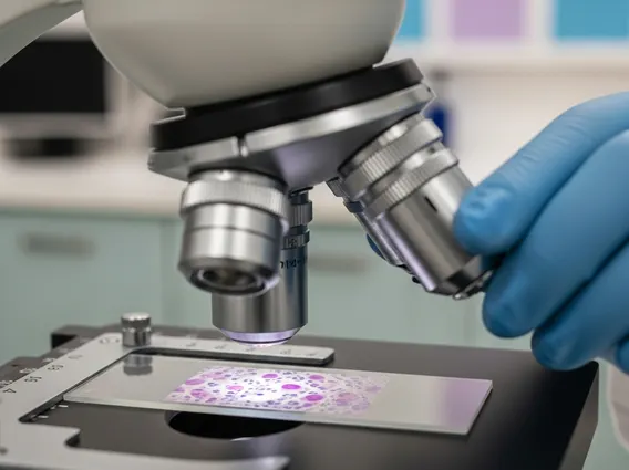
The process for mycosis fungoides patch diagnosis causes involves thorough evaluation. Diagnosis primarily relies on a skin biopsy, where a tissue sample from an affected patch is microscopically examined by a dermatopathologist. This examination seeks specific features, such as atypical T-lymphocytes within the epidermis (epidermotropism) and characteristic cellular infiltrates. Due to the subtle nature of early changes, multiple biopsies may be necessary to confirm the diagnosis. Advanced tests like immunohistochemistry and T-cell receptor gene rearrangement studies are often performed on biopsy samples to detect T-cell clonality, aiding differentiation from benign inflammatory conditions.
The precise causes of Mycosis Fungoides remain largely elusive. It is neither hereditary nor contagious. Research suggests a complex interplay of genetic predispositions, environmental exposures, and immune system irregularities. While no single cause has been definitively identified, potential contributing factors include chronic antigen stimulation, certain viral infections, and cumulative genetic mutations leading to malignant T-cell transformation.
Treatment Options for Mycosis Fungoides Patch
Managing mycosis fungoides patch treatment options aims to alleviate symptoms, prevent disease progression, and enhance quality of life, as a definitive cure for MF is not yet available. Treatment strategies are highly personalized, considering disease stage, extent of skin involvement, patient health, and preferences. For the patch stage, therapies are primarily skin-directed and less aggressive.
| Treatment Type | Description | Application |
|---|---|---|
| Topical Corticosteroids | Anti-inflammatory creams or ointments to reduce redness, itching, and scaling. | First-line for localized patches. |
| Topical Chemotherapy | Medications like mechlorethamine or carmustine applied directly to skin. | Targets malignant T-cells in the epidermis. |
| Phototherapy (UVB/PUVA) | Exposure to specific wavelengths of ultraviolet light, sometimes with a photosensitizing drug (psoralen). | Effective for widespread patches; requires careful monitoring. |
| Topical Retinoids | Gels or creams containing retinoids (e.g., bexarotene) to normalize skin cell growth. | Used to manage skin lesions. |
For patients with more extensive patch-stage disease or those unresponsive to initial therapies, systemic treatments might be considered. These are typically reserved for later stages or more aggressive presentations and can include oral retinoids, interferon-alpha, or low-dose methotrexate. Regular follow-up with a dermatologist or oncologist is essential to monitor disease activity and adjust treatment.