Mri Ultrasound Fusion Guided Biopsy
Mri Ultrasound Fusion Guided Biopsy is an advanced diagnostic procedure primarily used in the detection and characterization of prostate cancer. This innovative technique combines the strengths of two powerful imaging modalities to enhance diagnostic accuracy and precision.
Key Takeaways
- Mri Ultrasound Fusion Guided Biopsy combines detailed MRI scans with real-time ultrasound for highly accurate tissue sampling.
- It is primarily used for diagnosing prostate cancer, especially when initial screenings indicate potential issues.
- The procedure involves mapping suspicious areas from an MRI onto live ultrasound images to guide biopsy needles precisely.
- Key benefits include improved detection of clinically significant cancers, reduced need for repeat biopsies, and fewer complications.
- This method enhances diagnostic confidence by ensuring samples are taken from the most concerning areas.
What is MRI Ultrasound Fusion Guided Biopsy?
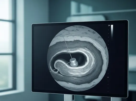
Mri Ultrasound Fusion Guided Biopsy is a sophisticated medical procedure that integrates magnetic resonance imaging (MRI) data with real-time ultrasound imaging during a biopsy. This fusion technology allows physicians to precisely target suspicious lesions that may not be visible on ultrasound alone, significantly improving the accuracy of tissue sampling. The fundamental principle behind what is Mri Ultrasound Fusion Guided Biopsy lies in leveraging the high-resolution anatomical detail provided by MRI to guide the biopsy needle with unparalleled precision.
Traditionally, prostate biopsies have relied solely on ultrasound guidance, which can sometimes miss significant cancers or lead to over-sampling of benign tissue. By fusing pre-biopsy MRI images, which are highly effective at identifying and characterizing suspicious areas within the prostate, with live ultrasound, clinicians gain a comprehensive, three-dimensional view. This integrated approach ensures that tissue samples are collected from the most concerning regions, thereby enhancing the diagnostic yield and reducing the likelihood of missing aggressive cancers.
How MRI Ultrasound Fusion Guided Biopsy Works
The process of Mri Ultrasound Fusion Guided Biopsy begins with a detailed multiparametric MRI of the prostate. This MRI scan identifies and maps any suspicious lesions, providing crucial information about their size, location, and characteristics. These MRI images are then loaded into specialized software that will be used during the biopsy procedure.
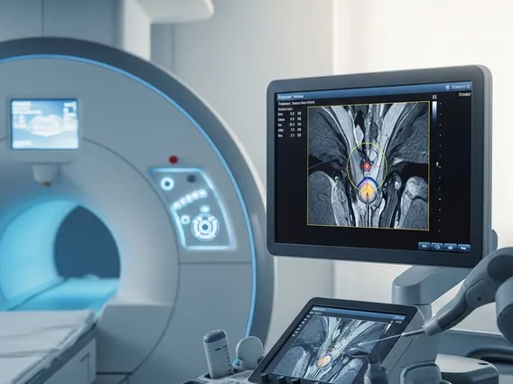
During the biopsy itself, the patient is positioned, and a real-time ultrasound probe is used to visualize the prostate. The core innovation of this technique, explaining How does MRI ultrasound fusion biopsy work?, is the fusion of these two imaging datasets. The pre-recorded MRI images are digitally overlaid and synchronized with the live ultrasound images. This creates a composite, real-time view where the physician can see both the detailed MRI findings and the current ultrasound anatomy simultaneously. This allows for highly accurate navigation to the target areas identified on the MRI.
The MRI ultrasound fusion biopsy procedure explained involves several key steps:
- MRI Acquisition: A detailed multiparametric MRI is performed to identify and characterize suspicious prostate lesions.
- Image Fusion: The MRI data is uploaded to a fusion biopsy system, which then merges it with real-time ultrasound images of the prostate.
- Targeting: The physician uses the fused images to precisely guide the biopsy needle to the identified suspicious areas.
- Tissue Sampling: Multiple tissue samples (cores) are taken from the targeted lesions, as well as from other areas of the prostate following a systematic approach.
- Pathological Analysis: The collected tissue samples are sent to a pathologist for microscopic examination to determine the presence and aggressiveness of cancer.
This precise targeting minimizes the number of biopsy cores needed while maximizing the chance of obtaining diagnostic tissue from the most relevant areas, leading to more confident and accurate diagnoses.
Benefits of MRI Ultrasound Fusion Guided Biopsy
The adoption of Mri Ultrasound Fusion Guided Biopsy offers several significant advantages over traditional, systematic ultrasound-guided biopsies. One of the primary Benefits of MRI ultrasound fusion biopsy is its significantly improved accuracy in detecting clinically significant prostate cancers. By directly targeting suspicious lesions identified on MRI, the procedure reduces the risk of missing aggressive cancers that might otherwise be overlooked by random sampling.
Studies have shown that MRI-ultrasound fusion biopsy can detect up to 30% more clinically significant prostate cancers compared to standard biopsy methods, as reported by various medical institutions and research (e.g., American Urological Association guidelines). This enhanced detection rate is crucial for timely and appropriate treatment planning. Furthermore, the precision of the fusion technique often leads to a reduction in the number of biopsy cores taken, which can decrease the risk of complications such as infection, bleeding, and discomfort for the patient.
In addition to improved diagnostic accuracy and reduced complications, this advanced biopsy method can also help in risk stratification. By accurately identifying the grade and extent of cancer, physicians can make more informed decisions regarding active surveillance, surgery, or radiation therapy. This personalized approach to diagnosis and treatment ultimately contributes to better patient outcomes and a more efficient use of healthcare resources.