Mri Guided Biopsy
MRI-guided biopsy is a sophisticated medical procedure that utilizes advanced imaging technology to precisely locate and sample suspicious tissues within the body. This technique is particularly valuable for areas not clearly visible through other imaging methods, ensuring accurate diagnosis and guiding subsequent treatment decisions.
Key Takeaways
- MRI-guided biopsy uses Magnetic Resonance Imaging to pinpoint and sample abnormal tissue.
- It offers high precision for lesions not detectable by ultrasound or mammography.
- The procedure involves real-time MRI guidance to ensure accurate tissue collection.
- Benefits include improved diagnostic accuracy, reduced need for more invasive surgeries, and better treatment planning.
- It is commonly used for suspicious findings in the breast, prostate, and other soft tissues.
What is MRI Guided Biopsy?
MRI-guided biopsy refers to a minimally invasive diagnostic procedure that employs Magnetic Resonance Imaging (MRI) to guide a biopsy needle to a specific area of concern within the body. This method is crucial when a suspicious lesion is identified on an MRI scan but cannot be visualized or accurately targeted using other imaging techniques, such as ultrasound or mammography. The primary goal is to obtain a tissue sample (biopsy) from the abnormal area for pathological examination, which helps determine if the cells are benign or malignant.
The procedure leverages the detailed, high-resolution images produced by MRI, which can differentiate between various soft tissues and highlight abnormalities that might be subtle or deeply located. This precision is vital for diagnosing conditions like certain types of breast cancer, prostate cancer, or lesions in other soft tissues that require a definitive diagnosis for appropriate medical management. According to the American Cancer Society, MRI can detect additional cancers in high-risk women that mammography might miss, often necessitating an MRI-guided biopsy for confirmation.
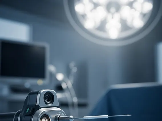
How Does MRI Guided Biopsy Work?
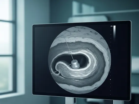
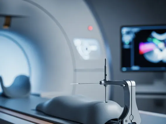
The process of an MRI guided biopsy procedure explained involves several careful steps to ensure accuracy and patient safety. Initially, the patient lies face down on a special MRI-compatible table, which is then moved into the MRI scanner. For breast biopsies, the breast is positioned in a coil with openings that allow access for the biopsy. Multiple MRI images are taken to precisely locate the target lesion in three dimensions.
Once the suspicious area is identified, the radiologist uses a computer workstation to plan the needle trajectory. A small incision is made in the skin after administering a local anesthetic to numb the area. Using real-time MRI guidance, a specialized biopsy needle is advanced through the skin and tissue to the exact location of the lesion. The MRI scanner takes additional images to confirm the needle’s position. Multiple tissue samples are then collected using a vacuum-assisted device or core needle biopsy system. After sufficient samples are obtained, a small marker clip may be placed at the biopsy site to facilitate future follow-up imaging or surgical procedures. The needle is removed, and pressure is applied to the site to minimize bleeding. The entire procedure typically takes about 30 to 60 minutes, followed by a brief recovery period.
The precision offered by MRI guidance significantly enhances the likelihood of obtaining diagnostic tissue from the target lesion, reducing the need for repeat biopsies or more invasive surgical procedures. This method is particularly beneficial for lesions that are small, deep, or only visible on MRI.
Benefits of MRI Guided Biopsy
MRI-guided biopsy offers several significant advantages, particularly in the diagnosis of complex or elusive lesions. Its primary benefit lies in its exceptional accuracy, allowing clinicians to target abnormalities that might be missed or are difficult to access with other imaging modalities. This precision translates into a higher diagnostic yield, meaning a greater chance of obtaining a definitive diagnosis from the initial biopsy.
Key benefits include:
- Enhanced Accuracy: MRI provides superior soft tissue contrast, enabling the visualization of lesions not seen on mammography or ultrasound, leading to more precise targeting.
- Reduced Invasiveness: As a minimally invasive procedure, it typically involves a small skin incision, leading to less pain, scarring, and a quicker recovery compared to open surgical biopsies.
- Early Detection: For certain conditions, such as some types of breast cancer in high-risk individuals or prostate cancer, MRI-guided biopsy can detect and diagnose cancers at an earlier stage, potentially improving treatment outcomes.
- Improved Treatment Planning: A precise diagnosis obtained through MRI-guided biopsy allows for more informed and tailored treatment strategies, avoiding unnecessary interventions or ensuring appropriate therapy.
- Patient Comfort: While the procedure requires remaining still in the MRI scanner, it often avoids the discomfort and risks associated with more extensive surgical interventions.
The ability of MRI-guided biopsy to accurately sample suspicious areas, especially in challenging anatomical locations, makes it an invaluable tool in modern diagnostic medicine. Its role continues to expand, particularly in oncology, where accurate tissue diagnosis is paramount for effective patient care.