Mrcp
Magnetic Resonance Cholangiopancreatography (MRCP) is a non-invasive medical imaging technique that uses a powerful magnetic field and radio waves to create detailed images of the bile ducts, pancreatic duct, and gallbladder.
Key Takeaways
- MRCP is a non-invasive MRI technique for visualizing bile and pancreatic ducts.
- It helps diagnose conditions like gallstones, tumors, inflammation, and strictures without radiation.
- The procedure involves lying still in an MRI scanner, often after fasting, and typically takes 30-60 minutes.
- MRCP is highly accurate for detecting obstructions and abnormalities in the biliary and pancreatic systems.
- Results are interpreted by a radiologist and guide further medical management.
What is MRCP (Magnetic Resonance Cholangiopancreatography)?
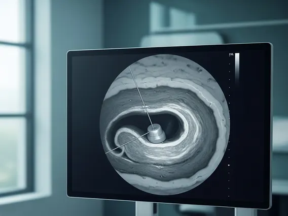
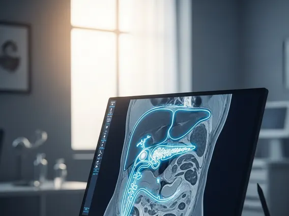
MRCP (Magnetic Resonance Cholangiopancreatography) is a specialized type of magnetic resonance imaging (MRI) that provides detailed images of the biliary and pancreatic systems. Unlike traditional X-rays or CT scans, MRCP does not use ionizing radiation, making it a safer option for repeated examinations or for patients sensitive to radiation. This advanced imaging technique focuses specifically on fluid-filled structures, such as the bile ducts within the liver, the common bile duct, the gallbladder, and the pancreatic duct, making them appear bright against the darker background of surrounding tissues.
The primary purpose of an MRCP medical procedure is to diagnose and evaluate a range of conditions affecting these vital digestive pathways. It is particularly effective in identifying blockages, inflammation, and other abnormalities that can lead to symptoms like abdominal pain, jaundice, or unexplained weight loss. Its non-invasive nature and high resolution make it an invaluable tool in modern gastroenterology and hepatology.
Uses and Procedure of MRCP Scan
An MRCP scan uses and benefits are extensive, primarily revolving around its ability to visualize the complex anatomy of the biliary and pancreatic systems without the need for endoscopic intervention. It is frequently employed to detect gallstones, which can obstruct bile flow, as well as to identify tumors, cysts, or inflammation (such as pancreatitis or cholangitis). Furthermore, MRCP can assess for congenital anomalies, strictures (narrowing of ducts), and fluid collections.
The procedure for how an MRCP scan is performed is straightforward and typically takes between 30 to 60 minutes. Patients are usually asked to fast for several hours before the scan to reduce fluid in the stomach and duodenum, which can interfere with image clarity. During the scan, the patient lies on a movable table that slides into the large, tube-shaped MRI scanner. It is crucial for the patient to remain still throughout the examination to ensure clear images. In some cases, a contrast agent may be administered intravenously to enhance certain structures, though this is less common for standard MRCP which relies on the natural fluid within the ducts. The machine generates detailed cross-sectional images that a radiologist later interprets.
Clinical studies indicate that MRCP has a high diagnostic accuracy, often exceeding 90%, for detecting choledocholithiasis (bile duct stones) and other biliary obstructions, making it a highly reliable diagnostic tool.
Interpreting MRCP Scan Results
The mrcp results interpretation guide is essential for clinicians to understand the findings and determine the appropriate course of action for the patient. After the scan, a specialized radiologist meticulously reviews the hundreds of images generated. They look for several key indicators, including the presence, size, and location of stones within the bile or pancreatic ducts, any signs of inflammation or swelling, and the presence of strictures or dilations (widening) of the ducts. The radiologist also assesses for masses or tumors that might be obstructing the flow of bile or pancreatic fluid.
Normal MRCP results typically show clear, unobstructed bile and pancreatic ducts of appropriate caliber, with no visible stones, masses, or signs of inflammation. Abnormal findings, however, can point to various conditions, such as choledocholithiasis (stones in the common bile duct), cholangitis (inflammation of the bile ducts), pancreatitis (inflammation of the pancreas), or even pancreatic or bile duct cancers. The radiologist compiles these findings into a detailed report, which is then sent to the referring physician. The physician will discuss these results with the patient, correlating them with their symptoms, physical examination, and other diagnostic tests to formulate a comprehensive treatment plan.