Mixed Glioma
Mixed glioma represents a complex category of primary brain tumors characterized by the presence of more than one type of glial cell. Understanding this condition is crucial for accurate diagnosis and effective management.

Key Takeaways
- Mixed Glioma is a brain tumor containing a combination of different glial cell types, most commonly oligodendroglioma and astrocytoma components.
- Symptoms vary widely depending on tumor location and size, often including headaches, seizures, and neurological deficits.
- Diagnosis involves imaging (MRI) and tissue biopsy, which is essential for determining the specific cellular composition.
- Treatment typically involves a multi-modal approach, including surgery, radiation therapy, and chemotherapy.
- Prognosis is highly variable, influenced by factors such as tumor grade, molecular markers, and the patient’s overall health.
What is Mixed Glioma?
Mixed Glioma refers to a type of primary brain tumor that exhibits features of more than one glial cell lineage. Historically, these tumors were often termed “oligoastrocytomas” due to their perceived mixture of oligodendroglial and astrocytic components. However, advancements in molecular pathology have refined our understanding, revealing that many tumors previously classified as mixed gliomas are predominantly either astrocytic or oligodendroglial with specific genetic mutations. The World Health Organization (WHO) Classification of Tumors of the Central Nervous System now emphasizes molecular markers, such as 1p/19q co-deletion and IDH mutations, which are critical for precise classification and guiding treatment decisions.
These tumors originate from glial cells, which are the supportive cells of the brain and spinal cord. Their mixed cellular composition makes them distinct from pure astrocytomas or oligodendrogliomas, although the exact proportion of each cell type can vary significantly. The grade of a mixed glioma, ranging from Grade II (low-grade) to Grade III (anaplastic), is determined by its cellular features and mitotic activity, directly impacting its biological behavior and treatment strategy.
Mixed Glioma Symptoms and Treatment Options
The presentation of mixed glioma symptoms can be diverse, largely depending on the tumor’s location, size, and growth rate within the brain. Common symptoms often include persistent headaches, which may worsen over time, and seizures, which can be the initial sign in many patients. Other neurological deficits may arise as the tumor grows and exerts pressure on specific brain regions. These can manifest as:
- Weakness or numbness on one side of the body
- Changes in personality or behavior
- Difficulties with speech or language (aphasia)
- Problems with vision or balance
- Cognitive impairments, such as memory loss or confusion
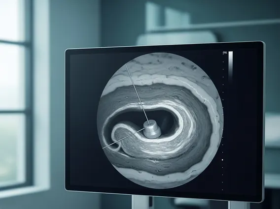
Diagnosis typically begins with imaging studies, such as magnetic resonance imaging (MRI), which can identify the tumor’s presence and characteristics. However, a definitive diagnosis requires a biopsy, where a tissue sample is surgically removed and examined under a microscope by a neuropathologist. This pathological analysis, combined with molecular testing, is crucial for accurate classification and grading.
Mixed glioma treatment options are tailored to the individual patient, considering factors like tumor grade, location, molecular profile, and the patient’s overall health. The primary treatment approach often involves:
| Treatment Modality | Description |
|---|---|
| Surgical Resection | Aims to remove as much of the tumor as safely possible without causing significant neurological damage. Maximal safe resection is often associated with improved outcomes. |
| Radiation Therapy | Uses high-energy rays to destroy cancer cells and prevent tumor recurrence, typically administered after surgery. |
| Chemotherapy | Utilizes drugs to kill cancer cells, often used in conjunction with radiation therapy, especially for higher-grade tumors or those with specific molecular markers (e.g., 1p/19q co-deletion). |
The combination and sequence of these treatments are determined by a multidisciplinary team of specialists, including neurosurgeons, neuro-oncologists, and radiation oncologists.
Prognosis and Long-Term Outlook for Mixed Glioma
The mixed glioma prognosis is highly variable and depends on several critical factors. Key prognostic indicators include the tumor’s WHO grade (Grade II vs. Grade III), the extent of surgical resection, and the presence of specific molecular markers. Tumors with IDH mutations and 1p/19q co-deletion, often classified as oligodendrogliomas, generally have a more favorable prognosis compared to those without these markers. For instance, according to data from the Central Brain Tumor Registry of the United States (CBTRUS), the median survival for adult patients with anaplastic oligodendroglioma (a type often previously grouped under mixed glioma) can be significantly longer than for anaplastic astrocytoma.
Long-term outlook also considers the patient’s age at diagnosis, performance status, and response to treatment. While complete cures are rare for most high-grade gliomas, advancements in treatment, particularly targeted therapies based on molecular profiles, continue to improve patient outcomes and quality of life. Regular follow-up imaging and clinical evaluations are essential to monitor for recurrence and manage any long-term side effects of treatment. Ongoing research aims to identify new therapeutic targets and strategies to further enhance the prognosis for individuals diagnosed with mixed glioma.