Microscopic Description
Microscopic Description is a fundamental practice in medicine, particularly in pathology, involving the detailed examination of biological samples at a cellular level. This process is critical for understanding disease processes and making accurate diagnoses.

Key Takeaways
- Microscopic Description involves the detailed examination of tissue or cell samples under a microscope to identify abnormalities.
- It is a cornerstone of medical diagnosis, especially in pathology, for conditions like cancer, infections, and inflammatory diseases.
- Effective descriptions require precision, objectivity, and adherence to standardized terminology to accurately convey findings.
- Guidelines for writing include systematic observation of architectural patterns, cellular features, and specific structures.
- The accuracy and clarity of these descriptions are paramount for guiding treatment decisions and providing prognostic information for patients.
What is Microscopic Description?
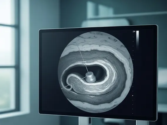
Microscopic Description refers to the detailed written account of observations made when examining biological specimens, such as tissue biopsies or cytology smears, under a microscope. This essential practice in diagnostic pathology involves pathologists meticulously analyzing cellular and architectural features to identify normal structures, disease processes, or abnormalities. The primary goal is to translate complex visual information into a clear, concise, and objective narrative that informs clinical decision-making.
This process typically begins after a specimen has been collected, processed, and stained, making various cellular components visible. Pathologists assess characteristics like cell size, shape, nuclear features, cytoplasmic details, tissue architecture, and the presence of any foreign elements or inflammatory cells. The resulting description forms a crucial part of the pathology report, providing the foundation for diagnosing a wide range of conditions, from infectious diseases to various forms of cancer.
Guidelines for Writing Microscopic Descriptions and Practical Examples
Writing an effective microscopic description requires a systematic approach, precision, and the use of standardized terminology to ensure clarity and reproducibility. Pathologists follow specific guidelines to accurately convey their observations, which are vital for other clinicians interpreting the report.
Key elements to include in a microscopic description are:
- Tissue Type and General Architecture: Identifying the organ or tissue origin and its overall structural arrangement.
- Cellularity: Assessing the density of cells within the tissue.
- Cellular Morphology: Describing individual cell characteristics, including nuclear size, shape, chromatin pattern, nucleoli, and cytoplasmic features.
- Architectural Patterns: Noting how cells are arranged (e.g., glands, nests, sheets, cords) and any deviations from normal.
- Stroma: Describing the supporting connective tissue and any changes within it (e.g., fibrosis, inflammation).
- Specific Features: Identifying the presence of inflammation, necrosis, mitosis, atypical cells, or other significant findings.
For example, when describing a biopsy from a suspected inflammatory condition, a pathologist might note: “Sections show colonic mucosa with preserved crypt architecture. There is a mild to moderate increase in chronic inflammatory cells (lymphocytes, plasma cells) in the lamina propria, without significant neutrophilic infiltration or cryptitis. No granulomas or dysplasia are identified.” This concise narrative provides a clear picture of the findings. Another example, for a tumor, could be: “Biopsy shows an infiltrative proliferation of atypical glandular structures lined by columnar cells with pseudostratified nuclei, prominent nucleoli, and moderate pleomorphism. Numerous mitotic figures are present. Desmoplastic stromal reaction is noted.” These microscopic description examples illustrate how specific observations are translated into a diagnostic narrative.
Importance of Microscopic Description in Medical Diagnosis
The importance of microscopic description in medical diagnosis cannot be overstated, as it serves as a cornerstone for accurate disease identification, prognosis, and treatment planning. Pathologists’ detailed observations under the microscope provide definitive diagnoses for a vast array of conditions, ranging from benign inflammatory processes to aggressive malignancies.
Microscopic descriptions are essential for:
- Accurate Diagnosis: Providing the definitive identification of diseases, such as differentiating between various types of cancer, identifying infectious agents, or characterizing inflammatory conditions. Without this detailed analysis, many diagnoses would remain speculative.
- Disease Staging and Grading: For cancers, the microscopic description helps determine the tumor type, grade (how aggressive it appears), and extent of invasion, all of which are critical for staging and guiding therapeutic strategies.
- Guiding Treatment Decisions: Clinicians rely heavily on the pathology report, which includes the microscopic description, to select the most appropriate treatment. For instance, identifying specific cellular features might indicate responsiveness to certain targeted therapies.
- Prognostic Information: The morphological features described can offer insights into the likely course of a disease and patient outcomes, helping clinicians counsel patients effectively.
According to the College of American Pathologists (CAP), pathology reports, which are heavily reliant on accurate microscopic descriptions, are integral to approximately 70% of all medical diagnoses, underscoring their profound impact on patient care. The meticulous work of pathologists in generating these descriptions ensures that patients receive precise diagnoses and optimal management plans, ultimately improving health outcomes.