Microcalcification
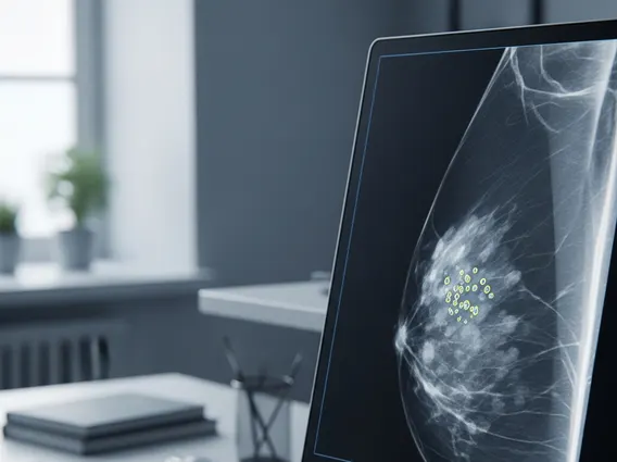
Microcalcifications are tiny calcium deposits that can appear in various tissues throughout the body, but are most commonly discussed in the context of breast health. Their presence is often detected during mammography, serving as an important indicator that may require further investigation.
Key Takeaways
- Microcalcifications are small calcium deposits, frequently found in breast tissue during mammograms.
- They are often benign, resulting from normal aging, inflammation, or non-cancerous conditions.
- However, certain patterns and shapes of microcalcifications can indicate a higher risk of breast cancer.
- Radiologists classify microcalcifications by their appearance and distribution to assess their potential significance.
- Further diagnostic steps, such as biopsy, may be necessary to determine if microcalcifications are benign or malignant.
What is Microcalcification? Definition and Underlying Causes
Microcalcification refers to minute calcium deposits that form within soft tissues, particularly in the breast. These deposits are too small to be felt during a physical exam and are typically identified on a mammogram as bright white spots. They are a common finding, especially in women over 50, and can result from various cellular processes, including inflammation, aging, or cellular necrosis.
The causes of microcalcifications are diverse, ranging from benign physiological changes to indicators of underlying pathology. Many are benign, often associated with:
- Aging: Normal wear and tear on breast tissue.
- Inflammation or Infection: Past mastitis or other inflammatory conditions.
- Fibrocystic Changes: Common non-cancerous breast conditions where calcium can deposit in cyst walls or stromal tissue.
- Trauma or Prior Surgery: Healing processes after injury or surgical procedures.
- Benign Tumors: Non-cancerous growths like fibroadenomas that can undergo calcification.
- Vascular Calcifications: Calcium deposits within blood vessel walls, distinct from those within breast tissue.
Crucially, microcalcifications can also be associated with precancerous conditions or early-stage breast cancer, such as ductal carcinoma in situ (DCIS). In these instances, the calcium deposits often form as a byproduct of rapid cell turnover, cellular debris, and necrosis within the abnormal or cancerous tissue, making their detection a vital part of early cancer diagnosis.
Types of Breast Microcalcifications and Their Cancer Risk
The appearance and distribution of types of breast microcalcifications are critical for radiologists in assessing their potential significance. Radiologists meticulously evaluate their shape, size, density, and pattern on a mammogram to distinguish between benign findings and those warranting further investigation.
The microcalcification breast cancer risk is directly correlated with these morphological features. Benign calcifications often appear scattered, round, or punctate, and are typically uniform, indicating a low likelihood of cancer. Malignant or suspicious calcifications, conversely, tend to be pleomorphic, fine linear, branching, or amorphous. Their distribution is also key; clustered or segmental patterns are generally more concerning than diffuse patterns. Early detection through mammography is paramount, as microcalcifications can be the earliest sign of breast cancer.
According to data from the American Cancer Society, while most breast microcalcifications are benign, approximately 10-20% of biopsies performed for suspicious microcalcifications ultimately reveal breast cancer. This underscores the importance of careful evaluation. (Source: American Cancer Society, general statistics on breast cancer detection).
To further clarify the distinction, here’s a summary of common types and their associated risk:
| Type of Microcalcification | Description | Associated Cancer Risk |
|---|---|---|
| Benign (Typically) | Round, punctate, coarse, vascular, rim-like, or eggshell-like; often scattered or diffuse. | Very low risk; usually requires no further action beyond routine screening. |
| Indeterminate (Potentially Suspicious) | Amorphous (indistinct shape), coarse heterogeneous (irregular, varying size); often grouped. | Moderate risk (5-20% chance of malignancy); often warrants a biopsy. |
| Suspicious (High Risk) | Fine linear, fine linear branching, pleomorphic (varying shapes/sizes); often clustered or segmental. | High risk (20-90% chance of malignancy); biopsy is strongly recommended. |
When microcalcifications are deemed suspicious, a stereotactic biopsy is typically performed for precise tissue sampling and pathological examination. This is essential for obtaining a definitive diagnosis and guiding subsequent treatment decisions if malignancy is confirmed. Regular mammographic screening remains the most effective tool for detecting these subtle changes early.