
Mediastinal Pleura
The mediastinal pleura is a vital component of the human respiratory system, forming part of the protective lining around the lungs. Understanding its structure and role is crucial for comprehending lung mechanics and the spread of thoracic diseases.
Key Takeaways
- The mediastinal pleura is a specific region of the parietal pleura, lining the lateral aspects of the mediastinum.
- Its primary role is to separate the lungs from the central mediastinal organs, preventing direct contact and friction.
- It contributes to the integrity of the pleural cavity, facilitating smooth lung expansion and contraction.
- Anatomically, it forms a crucial boundary, reflecting onto the visceral pleura at the lung hilum.
What is the Mediastinal Pleura?
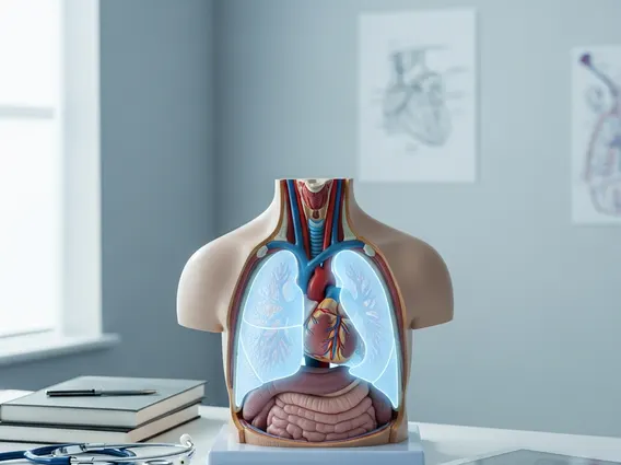
The mediastinal pleura definition refers to the portion of the parietal pleura that covers the lateral surfaces of the mediastinum. To understand what is mediastinal pleura, it’s essential to recognize it as one of the four main divisions of the parietal pleura, alongside the costal, diaphragmatic, and cervical (cupula) pleura. This delicate serous membrane acts as a protective barrier, separating the lungs from the vital structures housed within the mediastinum, such as the heart, great vessels, trachea, and esophagus. Its smooth, moist surface minimizes friction, allowing the lungs to expand and contract freely during respiration without impeding the function of adjacent organs.
The integrity of the mediastinal pleura is paramount for maintaining the distinct compartments within the thoracic cavity. Damage or inflammation to this specific pleural region can have significant clinical implications, potentially leading to conditions like pleurisy or the spread of infections between the lungs and the mediastinum. For instance, according to a review published in the Journal of Thoracic Disease, pleural effusions, which can sometimes involve the mediastinal pleura, are a common clinical problem affecting millions globally each year, highlighting the importance of this membrane’s health.
Anatomy and Location
The mediastinal pleura anatomy is characterized by its precise location and reflections within the chest cavity. It extends from the sternum anteriorly to the vertebral column posteriorly, covering the entire lateral aspect of the mediastinum on both the right and left sides. This pleural layer is continuous with the other parts of the parietal pleura at various points. Superiorly, it merges with the cervical pleura; inferiorly, it connects with the diaphragmatic pleura. Anteriorly and posteriorly, it joins the costal pleura, forming a complete lining for the thoracic cavity.
A key anatomical feature of the mediastinal pleura is its reflection onto the root of the lung, also known as the hilum. At this point, the parietal pleura folds back on itself to become the visceral pleura, which directly covers the lung surface. This reflection creates a sleeve-like structure around the bronchi, pulmonary arteries, and veins as they enter and exit the lung. The inferior extension of this reflection forms the pulmonary ligament, a double layer of pleura that stabilizes the lower lobe of the lung.
The structures adjacent to the mediastinal pleura include:
- Heart and Pericardium: Directly medial to the pleura.
- Great Vessels: Aorta, vena cavae, and pulmonary arteries/veins.
- Trachea and Main Bronchi: Central airways.
- Esophagus: Posterior to the trachea.
- Phrenic and Vagus Nerves: Important nerves traversing the mediastinum.
- Thymus Gland: In the anterior mediastinum, especially in younger individuals.
Key Functions of the Mediastinal Pleura
The primary mediastinal pleura function is to provide a smooth, lubricated surface that facilitates the movement of the lungs within the thoracic cavity. By lining the mediastinum, it creates a distinct boundary that prevents the lungs from adhering to the central mediastinal organs. This separation is critical for efficient respiration, as it allows the lungs to expand and contract without friction or impedance from the heart, great vessels, or other structures.
Beyond facilitating movement, the mediastinal pleura also plays a significant protective role. It acts as a barrier against the spread of infection or inflammation between the lungs and the mediastinum. For example, if an infection originates in the lung, the intact mediastinal pleura can help contain it, preventing its direct extension into the mediastinal space where vital organs reside. Conversely, mediastinal infections or tumors may be contained by this pleural layer, delaying their spread to the lung parenchyma. This compartmentalization is a fundamental aspect of thoracic anatomy, contributing to the overall resilience and functional integrity of the respiratory and cardiovascular systems.


