Magnetic Resonance Spectroscopic Imaging
Magnetic Resonance Spectroscopic Imaging is an advanced diagnostic tool that provides crucial insights into the biochemical composition of tissues. This non-invasive technique complements conventional anatomical imaging by revealing metabolic changes indicative of various medical conditions.
Key Takeaways
- Magnetic Resonance Spectroscopic Imaging (MRSI) is an advanced MRI technique that maps biochemical changes in tissues.
- It measures the concentrations of specific metabolites, offering insights into cellular function and metabolism.
- MRSI works by detecting unique spectral signatures of compounds like choline, creatine, and N-acetylaspartate.
- Its primary applications include oncology for tumor characterization, and neurology for diagnosing and monitoring brain disorders.
- This technique provides functional information beyond structural images, aiding in diagnosis, prognosis, and treatment planning.
What is Magnetic Resonance Spectroscopic Imaging (MRSI)?
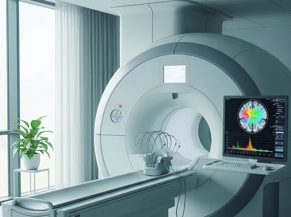
Magnetic Resonance Spectroscopic Imaging (MRSI) refers to a sophisticated, non-invasive medical imaging technique that combines the anatomical detail of magnetic resonance imaging (MRI) with the biochemical information derived from spectroscopy. It allows clinicians to visualize and quantify the distribution of various metabolites within specific regions of the body, offering a unique window into the metabolic state of tissues. Unlike standard MRI, which primarily provides structural images, MRSI generates metabolic maps that can highlight subtle biochemical alterations associated with disease processes. This advanced approach is often referred to as magnetic resonance spectroscopic imaging explained as a method for understanding tissue chemistry in vivo.
MRSI is particularly valuable because it can detect changes at a molecular level before structural abnormalities become apparent on conventional imaging. By analyzing the unique spectral “fingerprints” of different chemical compounds, MRSI provides functional information that aids in the early detection, characterization, and monitoring of numerous medical conditions. The technique is typically performed using the same MRI scanners, but with specialized hardware and software sequences designed to acquire spectroscopic data from multiple voxels (3D pixels) simultaneously.
How Magnetic Resonance Spectroscopic Imaging Works
Magnetic Resonance Spectroscopic Imaging operates on the same fundamental principles as conventional MRI but extends its capabilities to detect and quantify specific chemical compounds. The process begins with placing a patient in a strong magnetic field, which aligns the protons (hydrogen nuclei) within the body’s water and other molecules. Radiofrequency pulses are then applied, momentarily knocking these aligned protons out of alignment. When the pulses are turned off, the protons relax back into alignment, emitting radio signals in the process.
The key difference for MRSI is that it analyzes the subtle variations in these emitted signals based on the chemical environment of the protons. Different molecules, such as various metabolites, have protons that resonate at slightly different frequencies due to their surrounding electron clouds. This phenomenon, known as the chemical shift, allows MRSI to distinguish between and quantify various compounds. The signals are then processed to create a spectrum for each voxel, displaying peaks corresponding to different metabolites. The height and position of these peaks provide information about the concentration and type of metabolites present.
- N-acetylaspartate (NAA): A marker for neuronal viability and density, often decreased in neurodegenerative diseases.
- Choline (Cho): Associated with cell membrane turnover, often elevated in rapidly proliferating cells like tumors.
- Creatine (Cr): Involved in energy metabolism, often used as an internal reference for quantification due to its relatively stable concentration.
- Lactate (Lac): Indicates anaerobic metabolism, often elevated in areas of hypoxia or necrosis within tumors or stroke.
- Myo-inositol (mI): A glial cell marker, often elevated in certain neurological conditions.
Applications of Magnetic Resonance Spectroscopic Imaging
The applications of Magnetic Resonance Spectroscopic Imaging are diverse and continue to expand, particularly in the fields of oncology and neurology, where it offers critical diagnostic and prognostic information. In oncology, MRSI is invaluable for characterizing brain tumors, assessing their aggressiveness, and differentiating them from non-cancerous lesions or radiation necrosis. For instance, elevated choline levels and reduced NAA are often indicative of tumor presence and proliferation. It also plays a crucial role in monitoring treatment response, as changes in metabolite ratios can signal tumor regression or recurrence earlier than anatomical changes.
Beyond cancer, MRSI is increasingly utilized in the diagnosis and management of various neurological disorders. It can help in understanding the metabolic derangements in conditions such as Alzheimer’s disease, multiple sclerosis, stroke, and epilepsy. For example, reduced NAA in specific brain regions can indicate neuronal damage in neurodegenerative diseases, while elevated lactate may point to ischemic injury in stroke. The ability of MRSI to provide spatially resolved biochemical data makes it a powerful tool for guiding biopsies, planning radiation therapy, and evaluating the efficacy of therapeutic interventions. Its non-invasive nature and comprehensive metabolic insights position MRSI as a vital component in advanced clinical diagnostics and research.