Magnetic Resonance Angiography
Magnetic Resonance Angiography (MRA) is a non-invasive medical imaging technique that provides detailed images of blood vessels. It is a specialized application of Magnetic Resonance Imaging (MRI) used to detect, diagnose, and aid in the treatment of various vascular conditions.
Key Takeaways
- MRA is a non-invasive imaging technique for visualizing blood vessels.
- It uses strong magnetic fields and radio waves, not ionizing radiation.
- MRA helps diagnose conditions like aneurysms, blockages, and dissections.
- Contrast agents may be used to enhance image clarity.
- It offers detailed vascular information without surgical intervention.
What is Magnetic Resonance Angiography (MRA)?
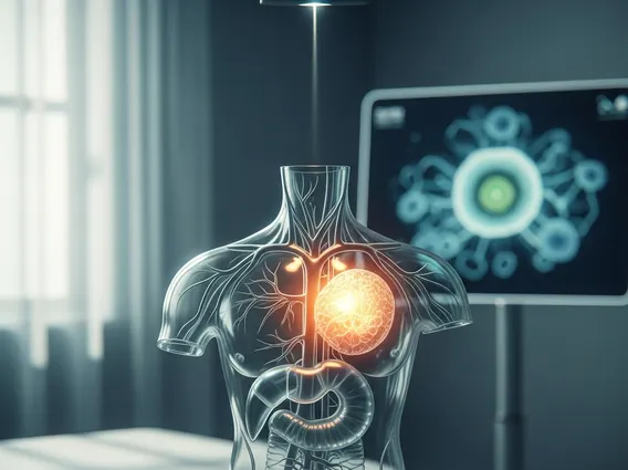
Magnetic Resonance Angiography (MRA) is a diagnostic imaging method that uses a powerful magnetic field, radio waves, and a computer to evaluate blood vessels and blood flow in the body. Unlike traditional angiography, MRA does not involve X-rays or ionizing radiation, making it a safer option for repeated examinations. It can produce detailed images of arteries and veins, helping clinicians identify abnormalities that could affect blood circulation. This advanced imaging technique is crucial for understanding what is Magnetic Resonance Angiography and its role in modern medicine, offering a clear view of vascular structures without invasive procedures.
MRA can visualize blood vessels in various parts of the body, including the brain, neck, heart, abdomen, and limbs. It is particularly effective at detecting issues such as narrowing (stenosis), blockages, aneurysms (bulges in vessel walls), and dissections (tears in vessel walls). The high-resolution images generated by MRA allow for precise diagnosis and treatment planning, often providing critical information that other imaging modalities might miss.
How Magnetic Resonance Angiography Works
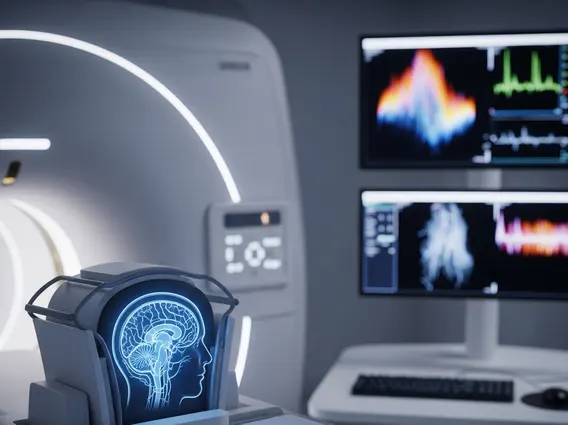
The fundamental principle behind Magnetic Resonance Angiography involves the use of a strong magnetic field to align the protons within the body’s water molecules. Radio waves are then briefly pulsed, knocking these aligned protons out of alignment. When the radio waves are turned off, the protons relax back into alignment, releasing energy signals that are detected by the MRA scanner. A computer processes these signals to create cross-sectional images of the body. For MRA, the technique is specifically adapted to highlight blood vessels.
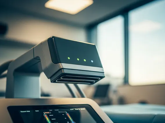
During a typical MRA procedure, the patient lies on a movable table that slides into a large, tunnel-like scanner. Depending on the area being examined, a contrast material, often gadolinium-based, may be injected intravenously. This contrast agent travels through the bloodstream, making the blood vessels appear brighter and more distinct on the MRA images, allowing for a clearer visualization of the vascular anatomy and any potential abnormalities. The entire procedure typically takes between 30 to 60 minutes, during which the patient must remain still. The scanner makes loud knocking noises, so earplugs or headphones are often provided. The detailed images produced help doctors assess blood flow and identify vascular issues.
Uses and Benefits of MRA Scans
MRA scan uses and benefits are extensive, making it an invaluable tool in diagnosing and managing a wide range of vascular conditions. Its non-invasive nature and ability to provide highly detailed images of blood vessels without radiation exposure are significant advantages. MRA is frequently used to:
- Detect and characterize aneurysms, particularly in the brain and aorta.
- Identify arterial blockages or narrowing (stenosis) that can lead to stroke or peripheral artery disease.
- Evaluate congenital abnormalities of blood vessels.
- Assess the extent of atherosclerotic disease.
- Plan surgical or interventional procedures, such as angioplasty or stent placement.
- Monitor the effectiveness of treatments for vascular conditions.
According to the American College of Radiology, MRA is a preferred imaging modality for many vascular assessments due to its excellent soft tissue contrast and ability to visualize blood flow without the risks associated with ionizing radiation. For instance, MRA of the carotid arteries is commonly performed to assess stroke risk by identifying significant narrowing. The benefits include its non-invasiveness, high diagnostic accuracy, and the absence of radiation, which is particularly important for patients requiring multiple follow-up scans.