
Intravenous Pyelography
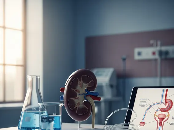
Intravenous Pyelography (IVP) is a diagnostic imaging test that uses X-rays and a special contrast dye to visualize the kidneys, ureters, and bladder. This procedure helps medical professionals assess the structure and function of the urinary tract.
Key Takeaways
- Intravenous Pyelography (IVP) is an X-ray imaging test that uses an injected contrast dye to highlight the urinary system.
- It helps diagnose conditions affecting the kidneys, ureters, and bladder, such as stones, tumors, or blockages.
- The procedure involves injecting a contrast agent into a vein, followed by a series of X-ray images as the dye travels through the urinary tract.
- While effective, IVP carries potential risks, including allergic reactions to the contrast dye and radiation exposure.
- Newer imaging techniques like CT and MRI have largely replaced IVP for many diagnostic purposes, though it remains relevant in specific clinical scenarios.
What is Intravenous Pyelography (IVP)?
Intravenous Pyelography (IVP) is a radiological examination that uses a contrast material injected into a vein to produce images of the kidneys, ureters, and bladder. This diagnostic tool allows healthcare providers to observe how the kidneys filter the contrast dye from the blood and how it flows through the urinary tract. The contrast agent makes these structures visible on X-ray images, revealing their size, shape, and position.
The purpose of intravenous pyelogram is primarily to detect and evaluate abnormalities within the urinary system. It can help identify conditions such as kidney stones, tumors, cysts, blockages, and congenital malformations. By visualizing the entire urinary pathway, IVP provides crucial information for diagnosing the cause of symptoms like flank pain, blood in the urine, or recurrent urinary tract infections.
The Intravenous Pyelography (IVP) Procedure Explained
The Intravenous Pyelography procedure typically begins with preparation, which may include fasting for several hours before the exam and sometimes taking a laxative to clear the bowels, ensuring clearer X-ray images. Upon arrival, the patient will lie on an X-ray table. A healthcare professional will then inject an iodine-based contrast dye into a vein, usually in the arm.
As the contrast dye circulates through the bloodstream, it is filtered by the kidneys and excreted into the urinary tract. A series of X-ray images are taken at specific intervals—typically a few minutes after injection, then at 5, 10, and 15 minutes, and sometimes later—to capture the dye as it fills the renal calyces, renal pelvis, ureters, and bladder. Compression may be applied to the abdomen during some images to help distend the renal collecting system. The entire procedure usually takes about 30 to 60 minutes, though it can sometimes be longer if delayed images are required.
Uses and Potential Risks of IVP Scans
IVP scans have historically been a valuable tool for diagnosing a range of urinary tract conditions. While its use has diminished with the advent of advanced imaging techniques like CT and MRI, it still holds specific indications. Common uses for IVP include:
- Detecting kidney stones (nephrolithiasis) and assessing their location and impact on urine flow.
- Identifying blockages in the urinary tract, such as those caused by tumors, strictures, or blood clots.
- Evaluating the extent of trauma to the kidneys or ureters.
- Diagnosing congenital abnormalities of the urinary system.
- Assessing the cause of recurrent urinary tract infections or unexplained flank pain.
Despite its diagnostic utility, IVP scans carry potential risks. The primary concern is an allergic reaction to the iodine-based contrast dye, which can range from mild symptoms like hives or nausea to severe reactions such as difficulty breathing or anaphylaxis. Patients with a history of allergies, asthma, or previous reactions to contrast agents are at higher risk. Another risk is kidney damage, particularly in individuals with pre-existing kidney disease, diabetes, or dehydration, as the contrast dye can be nephrotoxic. Additionally, IVP involves exposure to ionizing radiation, which, while generally low for a single procedure, is a cumulative risk over a lifetime. Due to these risks and the availability of alternative imaging methods, the decision to perform an IVP is carefully weighed by healthcare providers.


