Intracutaneous
Intracutaneous administration refers to a method of introducing substances directly into the dermis, the layer of skin located between the epidermis and the subcutaneous tissue. This technique is primarily used for diagnostic testing and certain vaccinations due to the unique immunological properties of the dermis.
Key Takeaways
- Intracutaneous administration involves injecting substances into the dermis, creating a characteristic wheal.
- It is distinct from subcutaneous injections, which target the fatty layer beneath the dermis, differing in depth, needle angle, and absorption rate.
- This method is crucial for diagnostic tests like the tuberculin skin test and allergy testing, leveraging the dermis’s immune cell concentration.
- The slow absorption from the dermal layer allows for localized reactions, making it ideal for observing immune responses.
- Proper technique is vital to ensure accurate test results and minimize discomfort for the patient.
What is Intracutaneous Administration?
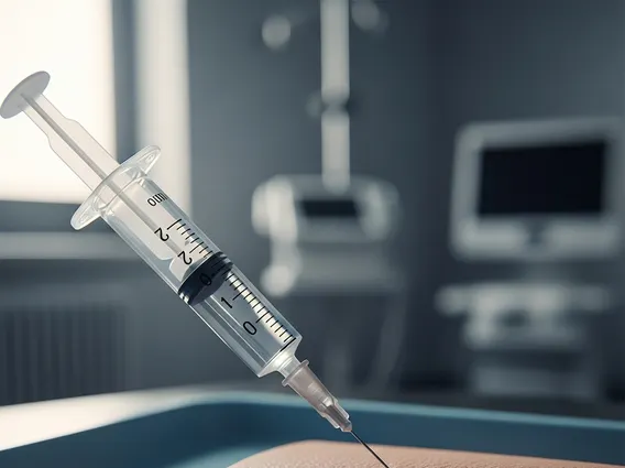
Intracutaneous administration, often referred to as an intradermal injection, is a precise method of delivering a substance into the dermis, the vascular layer of skin just below the epidermis. The primary goal of this technique is to introduce a small volume of fluid (typically 0.01 to 0.1 mL) into this specific layer, where it forms a distinct, raised blister-like area known as a wheal or bleb. The dermis is rich in immune cells, making it an ideal site for observing localized immune responses, which is why the intracutaneous injection meaning is often tied to diagnostic purposes.
The procedure involves inserting a short, fine-gauge needle at a very shallow angle, usually 5 to 15 degrees, almost parallel to the skin surface. The bevel of the needle should be visible through the epidermis as the injection is administered. The slow absorption rate from the dermis, compared to other injection routes, allows for a prolonged local effect, which is critical for the interpretation of skin tests. This method is less commonly used for systemic drug delivery due to the small volumes that can be administered and the slow absorption.
Intracutaneous vs. Subcutaneous Injections
Understanding the differences between intracutaneous vs subcutaneous injections is crucial for healthcare professionals, as each route serves distinct purposes and requires different techniques. While both involve injecting into the skin, they target different layers, leading to variations in absorption rates, needle angles, and common applications. The primary distinction lies in the depth of injection.
Intracutaneous injections target the dermis, a relatively thin layer, whereas subcutaneous injections deliver substances into the adipose (fatty) tissue located beneath the dermis. This deeper placement in subcutaneous tissue allows for larger volumes of medication to be administered and generally results in a slower, more sustained absorption into the bloodstream compared to intramuscular injections, but faster than intracutaneous absorption. The table below highlights key differences:
| Feature | Intracutaneous Injection | Subcutaneous Injection |
|---|---|---|
| Target Layer | Dermis (just below epidermis) | Subcutaneous tissue (fatty layer below dermis) |
| Needle Angle | 5-15 degrees | 45-90 degrees (depending on tissue amount) |
| Volume Administered | Very small (0.01-0.1 mL) | Small to moderate (0.5-2 mL) |
| Absorption Rate | Slowest (localized effect) | Slow to moderate (sustained systemic effect) |
| Common Uses | Diagnostic tests (e.g., TB, allergy), some vaccines | Insulin, heparin, some vaccines, pain medications |
| Characteristic Sign | Formation of a wheal/bleb | No wheal formation |
Common Intracutaneous Tests and Their Uses
The unique properties of intracutaneous administration make it invaluable for various diagnostic procedures. The intracutaneous test definition centers on introducing an antigen or allergen into the dermis to elicit a localized immune response, which can then be observed and measured. These tests rely on the high concentration of immune cells, such as Langerhans cells and T-lymphocytes, present in the dermal layer.
One of the most well-known applications is the tuberculin skin test, also known as the Mantoux test, used to screen for tuberculosis infection. A small amount of tuberculin purified protein derivative (PPD) is injected intracutaneously. A positive reaction, indicated by an area of induration (hardening) measured 48-72 hours later, suggests exposure to tuberculosis bacteria. According to the World Health Organization (WHO), tuberculin skin testing remains a vital tool in TB surveillance and diagnosis, particularly in high-burden countries.
Another common use is in allergy testing. During skin prick or intradermal allergy tests, tiny amounts of suspected allergens are injected intracutaneously. The presence and size of a localized wheal and flare reaction indicate sensitivity to specific allergens. This method helps identify triggers for conditions such as allergic rhinitis, asthma, and food allergies, guiding treatment and avoidance strategies. These tests provide specific, localized information that is critical for accurate diagnosis and patient management.