
Intestinal Villi
Intestinal Villi are microscopic, finger-like projections that line the inner surface of the small intestine, playing a crucial role in the digestive system. These specialized structures are essential for the efficient absorption of nutrients from digested food into the bloodstream.
Key Takeaways
- Intestinal Villi are tiny, finger-like projections found in the small intestine, significantly increasing its surface area.
- Their primary role is to facilitate the absorption of digested nutrients into the circulatory and lymphatic systems.
- Each villus contains a rich network of blood capillaries and a central lymphatic vessel (lacteal) to transport absorbed substances.
- The extensive surface area provided by villi and microvilli ensures highly efficient nutrient uptake.
- Proper functioning of Intestinal Villi is vital for overall nutrient assimilation and preventing malnutrition.
What are Intestinal Villi?
Intestinal Villi are small, numerous, finger-like projections that extend into the lumen of the small intestine. These structures are fundamental components of the digestive tract, designed to maximize the efficiency of nutrient uptake. Their presence dramatically increases the internal surface area of the small intestine, which is critical for absorbing the vast array of nutrients derived from the food we consume. Without these specialized structures, the body’s ability to absorb essential vitamins, minerals, carbohydrates, fats, and proteins would be severely compromised, leading to nutritional deficiencies.
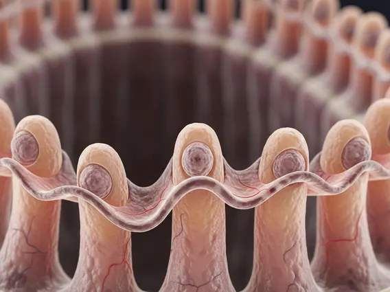
Structure and Key Functions of Intestinal Villi
The intricate structure of Intestinal Villi is perfectly adapted for their absorptive role. Each villus is covered by a layer of epithelial cells, primarily enterocytes, which themselves have microscopic projections called microvilli on their apical surface. This combined folding (villi and microvilli) creates an enormous surface area—estimated to be comparable to a tennis court—for nutrient absorption. Beneath the epithelial layer, each villus contains a dense network of blood capillaries and a central lymphatic vessel known as a lacteal.
The primary function of Intestinal Villi is to absorb digested nutrients. The capillaries within the villi absorb amino acids (from protein digestion) and monosaccharides (from carbohydrate digestion), transporting them directly to the liver via the portal vein. The lacteal, on the other hand, is responsible for absorbing dietary fats and fat-soluble vitamins, which are then transported into the lymphatic system before eventually entering the bloodstream. This dual absorption pathway ensures that all types of nutrients are efficiently processed and distributed throughout the body.
Key components within each Intestinal Villus include:
- Enterocytes: Epithelial cells forming the outer layer, equipped with microvilli to further increase surface area.
- Goblet Cells: Interspersed among enterocytes, these cells secrete mucus to lubricate and protect the intestinal lining.
- Capillary Network: A rich supply of blood vessels that absorb water-soluble nutrients.
- Lacteal: A central lymphatic vessel responsible for absorbing fats and fat-soluble vitamins.
- Lamina Propria: Connective tissue supporting the epithelial cells, containing immune cells.
Intestinal Villi and Nutrient Absorption
The process of Intestinal Villi absorption is highly efficient due to the vast surface area and the specialized cells lining the villi. As chyme (partially digested food) passes through the small intestine, nutrients come into direct contact with the villi. Enzymes present on the brush border of the enterocytes (known as brush border enzymes) complete the final stages of digestion, breaking down complex molecules into absorbable units.
For instance, carbohydrates are broken down into simple sugars like glucose, which are then actively transported into the enterocytes and subsequently into the capillaries. Proteins are digested into amino acids and small peptides, which follow a similar absorption pathway into the bloodstream. Fats, after being emulsified by bile and broken down by lipases, form micelles that are absorbed into enterocytes, re-esterified, and then packaged into chylomicrons before entering the lacteals. This intricate and coordinated process ensures that the body receives the necessary building blocks and energy sources to maintain health and function.


