Interval Breast Cancer
Interval Breast Cancer refers to a breast cancer detected between scheduled mammography screenings, often presenting with noticeable symptoms. Understanding this condition is crucial for timely diagnosis and effective management, as it can differ from cancers found during routine screening.
Key Takeaways
- Interval Breast Cancer is detected between routine mammograms, often due to new symptoms.
- These cancers can grow rapidly or be subtle, making them challenging to detect during screening.
- Common symptoms include new lumps, skin changes, or nipple discharge.
- Risk factors include dense breast tissue, younger age, and certain tumor characteristics.
- Diagnosis involves clinical examination, diagnostic imaging, and biopsy.
What is Interval Breast Cancer?
Interval Breast Cancer is a term used in oncology to describe a breast cancer that becomes clinically apparent after a negative screening mammogram but before the next scheduled screening. These cancers are significant because they are not detected by the routine screening process and often present due to patient-reported symptoms. They can represent either rapidly growing tumors that developed in the interval, or cancers that were present but missed on the previous mammogram due to factors like dense breast tissue or subtle imaging features. According to a study published in the journal Radiology, interval cancers account for 20-30% of all breast cancers diagnosed in screened populations, highlighting their clinical importance.
Symptoms, Causes, and Risk Factors for Interval Breast Cancer
Recognizing the signs of interval breast cancer is vital for early detection. The presentation of interval breast cancer symptoms causes patients to seek medical attention outside of their regular screening schedule. Common symptoms include the discovery of a new lump or mass in the breast, changes in breast size or shape, skin dimpling or puckering, nipple retraction, redness or scaling of the nipple or breast skin, and nipple discharge other than breast milk. These symptoms often prompt a diagnostic work-up.
The causes of interval breast cancer are multifactorial. Some interval cancers are genuinely new, rapidly growing tumors that develop quickly after a clear mammogram. Others are “missed” cancers, meaning they were present at the time of screening but were not identified due to factors such as:
- Tumor characteristics: Some cancers are less visible on mammograms, such as lobular carcinomas.
- Breast density: Dense breast tissue can obscure tumors, making them harder to detect.
- Radiological interpretation: Human error or the subtle nature of the lesion can lead to a missed diagnosis.
Several risk factors for interval breast cancer have been identified. These include having dense breast tissue, which reduces the sensitivity of mammography. Younger age at screening is also a risk factor, as younger women tend to have denser breasts and more aggressive tumor biology. Additionally, certain tumor characteristics, such as high histological grade, rapid proliferation rates, and specific molecular subtypes (e.g., triple-negative breast cancer), are associated with a higher likelihood of presenting as an interval cancer.
Diagnosing Interval Breast Cancer
Diagnosing Interval Breast Cancer typically begins when a patient or their physician notices a new breast change or symptom after a recent negative mammogram. The diagnostic process is thorough, aiming to confirm the presence of cancer and characterize it fully. This usually involves a combination of clinical examination, advanced imaging, and tissue sampling.
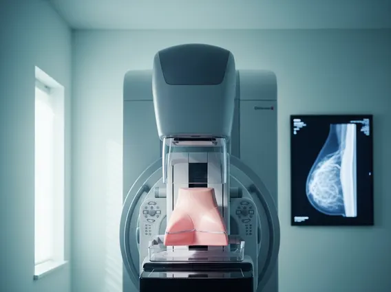
When a suspicious finding is noted, the initial steps often include a diagnostic mammogram, which provides more detailed views than a screening mammogram, often with spot compression and magnification. This is frequently followed by a breast ultrasound, which is particularly useful for evaluating palpable lumps and distinguishing between solid masses and fluid-filled cysts. Magnetic Resonance Imaging (MRI) of the breast may also be utilized, especially for women with dense breasts or when there is a strong suspicion of malignancy not fully characterized by mammography and ultrasound. These imaging modalities help to precisely locate the abnormality and assess its characteristics.
If imaging reveals a suspicious lesion, a biopsy is performed to obtain tissue for pathological examination. This is the definitive step in diagnosing breast cancer. Biopsy methods include fine-needle aspiration, core needle biopsy, or surgical biopsy, depending on the lesion’s size, location, and characteristics. The tissue samples are then analyzed by a pathologist to confirm the presence of cancer, determine its type, grade, and other important features, which guide subsequent treatment decisions. Comparing current imaging findings with previous mammograms is also a crucial step to understand why the cancer was not detected earlier and to inform future screening strategies.