Hydroureter
Hydroureter is a medical condition characterized by the dilation of the ureter, the tube that carries urine from the kidney to the bladder. This enlargement typically occurs due to an obstruction that prevents the normal flow of urine, leading to a buildup of pressure.

Key Takeaways
- Hydroureter involves the widening of the ureter, usually caused by a blockage in urine flow.
- It can result from various factors, including kidney stones, tumors, or anatomical abnormalities.
- Symptoms often include flank pain, frequent urination, and sometimes fever, depending on the cause.
- Diagnosis relies on imaging techniques like ultrasound, CT scans, and MRI.
- Treatment focuses on addressing the underlying cause to restore normal urine drainage and prevent kidney damage.
What is Hydroureter?
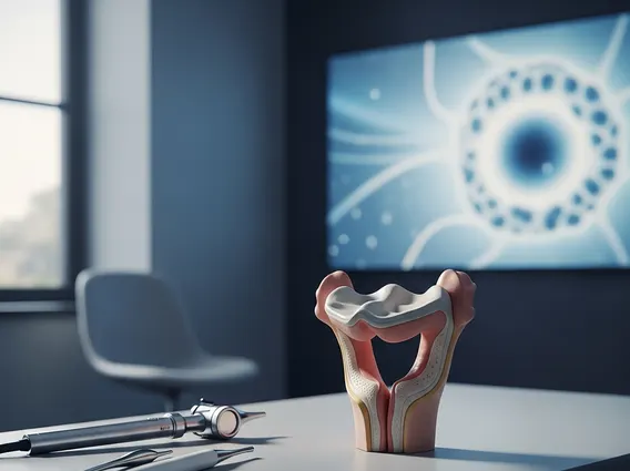
Hydroureter refers to the distension and dilation of the ureter, the tube that transports urine from the kidney to the urinary bladder. This condition arises when an obstruction along the urinary tract prevents normal urine flow, causing urine to back up and accumulate within the ureter. The increased pressure stretches and widens the ureteral wall. Untreated, this can lead to hydronephrosis (dilation of the renal pelvis and calyces) and impaired kidney function. It can be unilateral or bilateral, depending on the obstruction’s location.
Causes, Symptoms, and Diagnosis of Hydroureter
Understanding the factors contributing to hydroureter causes and symptoms is crucial. The primary cause is an obstruction impeding urine flow, which can be intrinsic (within the ureter) or extrinsic (compressing from outside).
Common causes include:
- Kidney stones (ureterolithiasis): A frequent cause, blocking the ureter.
- Tumors: Growths in the ureter, bladder, prostate, or adjacent organs.
- Strictures: Narrowing of the ureter from scar tissue.
- Congenital abnormalities: Structural issues like ureteropelvic junction (UPJ) obstruction.
- Pregnancy: The enlarging uterus can compress ureters, often on the right side.
Symptoms vary by cause and severity. Patients may experience:
- Flank pain: A dull ache or sharp pain in the side or back, potentially radiating to the groin.
- Frequent or painful urination.
- Nausea and vomiting.
- Fever and chills: Indicating a urinary tract infection.
- Hematuria: Blood in the urine.
Diagnosing hydroureter involves physical examination, medical history, and imaging. Initial assessment may include blood tests for kidney function and urinalysis.
Key diagnostic tools:
- Ultrasound: Non-invasive, often first-line to visualize the dilated ureter and kidney.
- Computed Tomography (CT) scan: Provides detailed images, identifying stones or tumors.
- Magnetic Resonance Imaging (MRI): Useful when radiation is a concern, offering soft tissue imaging.
- Cystoscopy: Visualizes the bladder and ureteral openings.
Hydroureter Treatment Options
The goal of hydroureter treatment options is to relieve the obstruction, restore normal urine flow, and prevent kidney damage. The specific approach depends on the underlying cause, severity, and patient health.
Treatment strategies may include:
- Observation: For mild, asymptomatic cases, such as physiological hydroureter during pregnancy.
- Medical Management: Pain relievers, antibiotics for infection, or alpha-blockers to aid stone passage.
- Interventional Procedures:
- Ureteral stent placement: A temporary tube to bypass obstruction.
- Nephrostomy tube placement: Drains urine directly from the kidney externally in severe cases.
- Lithotripsy: Techniques to break up kidney stones.
- Surgical Intervention: Endoscopic surgery (e.g., ureteroscopy) to remove stones or incise strictures, or open/laparoscopic surgery for complex issues like tumors or congenital anomalies.
Early diagnosis and appropriate treatment are vital to prevent complications such as kidney damage, recurrent infections, and chronic kidney disease.