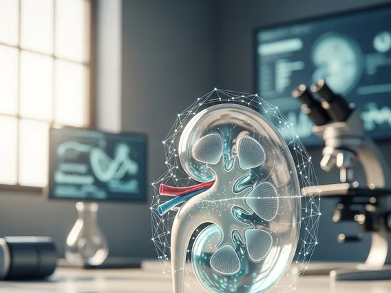
Hydronephrosis
Hydronephrosis is a medical condition characterized by the swelling of one or both kidneys due to a buildup of urine. This occurs when urine cannot drain from the kidney into the bladder, often due to a blockage or obstruction.
Key Takeaways
- Hydronephrosis involves kidney swelling from urine accumulation, often due to blockages in the urinary tract.
- Symptoms can range from flank pain and nausea to urinary issues, or the condition may be asymptomatic.
- Common causes include kidney stones, prostate enlargement, strictures, and congenital defects.
- Diagnosis relies on imaging tests such as ultrasound, CT scans, and MRI to identify the obstruction.
- Treatment focuses on relieving the obstruction and preserving kidney function, often involving drainage procedures or surgery.
What is Hydronephrosis?
Hydronephrosis refers to the distension and swelling of the renal pelvis and calyces, typically caused by an obstruction of urine flow. This condition can affect one kidney (unilateral hydronephrosis) or both kidneys (bilateral hydronephrosis), leading to increased pressure within the kidney. If left untreated, this sustained pressure can damage the kidney tissue, potentially leading to impaired kidney function or even kidney failure.
It is a relatively common condition, affecting individuals of all ages, from infants to the elderly. For instance, studies suggest that hydronephrosis is detected in approximately 1% of all pregnancies during prenatal ultrasound screenings, as reported by organizations like the American Urological Association.
Symptoms, Causes, and Diagnosis of Hydronephrosis
Recognizing the signs and understanding the origins of this condition are crucial for timely intervention. The hydronephrosis symptoms causes can vary significantly depending on the underlying obstruction, its severity, and whether the condition is acute or chronic. Some individuals may experience no symptoms, especially if the condition develops slowly. When symptoms do occur, they often include:
- Flank pain or back pain, which can be mild or severe
- Nausea and vomiting
- Frequent or urgent need to urinate
- Painful urination (dysuria)
- Blood in the urine (hematuria)
- Fever, if an infection is present
- Reduced urine output
Common causes of hydronephrosis include:
- Kidney stones: These are a frequent cause, blocking the ureter.
- Benign prostatic hyperplasia (BPH): An enlarged prostate in men can compress the urethra, obstructing urine flow.
- Tumors: Cancers in the kidney, bladder, prostate, or surrounding areas can cause blockages.
- Ureteral strictures: Narrowing of the ureters due to injury, infection, or previous surgery.
- Congenital abnormalities: Structural defects present at birth, such as a ureteropelvic junction (UPJ) obstruction.
- Vesicoureteral reflux (VUR): A condition where urine flows backward from the bladder into the ureters and kidneys.
Diagnosing hydronephrosis typically involves a combination of physical examination, medical history review, and various imaging tests. The primary goal of diagnosis is to identify the presence of kidney swelling and pinpoint the exact location and nature of the obstruction. Key diagnostic tools include ultrasound, which is often the first imaging test used, as well as Computed Tomography (CT) scans and Magnetic Resonance Imaging (MRI) for more detailed visualization. In some cases, an Intravenous Pyelogram (IVP) or CT Urogram, involving contrast dye, may be performed to visualize the urinary tract. These diagnostic methods help healthcare providers formulate an appropriate treatment plan.
Treatment Options for Hydronephrosis
The primary objective of hydronephrosis treatment options is to relieve the obstruction and restore normal urine flow, thereby preventing further kidney damage and preserving renal function. The specific treatment approach depends on the underlying cause, the severity of the hydronephrosis, and the patient’s overall health.
Common treatment strategies include:
- Drainage procedures: A ureteral stent may be placed to bypass the obstruction, or a nephrostomy tube can be inserted directly into the kidney to drain urine externally, especially in severe cases or with infection.
- Addressing the underlying cause: This may involve procedures for kidney stone removal (e.g., lithotripsy, ureteroscopy), surgical intervention to remove tumors or correct congenital abnormalities (e.g., pyeloplasty for UPJ obstruction), or medication to manage conditions like benign prostatic hyperplasia (BPH).
- Antibiotics: If a urinary tract infection (UTI) is present or suspected, antibiotics will be prescribed to clear the infection and prevent complications.
Regular monitoring of kidney function and follow-up imaging are essential after treatment to ensure the condition does not recur and that kidney health is maintained. Early diagnosis and appropriate treatment are vital for a positive outcome and to prevent long-term kidney damage.