Hydatidiform Mole
A hydatidiform mole, often referred to simply as a molar pregnancy, is a rare and abnormal form of pregnancy resulting from a genetic error during fertilization. This condition involves the growth of abnormal tissue within the uterus, rather than a viable embryo.
Key Takeaways
- A Hydatidiform Mole is an abnormal pregnancy where a non-viable embryo or no embryo develops, and the placenta grows into a mass of cysts.
- Symptoms often include vaginal bleeding, severe nausea, and unusually high human chorionic gonadotropin (hCG) levels.
- The primary cause is an error in the genetic material during fertilization, leading to either a complete or partial mole.
- Treatment involves surgical removal of the abnormal tissue, typically through dilation and curettage (D&C).
- Close follow-up with hCG monitoring is crucial after treatment to ensure complete resolution and detect any persistent disease.
What is a Hydatidiform Mole?
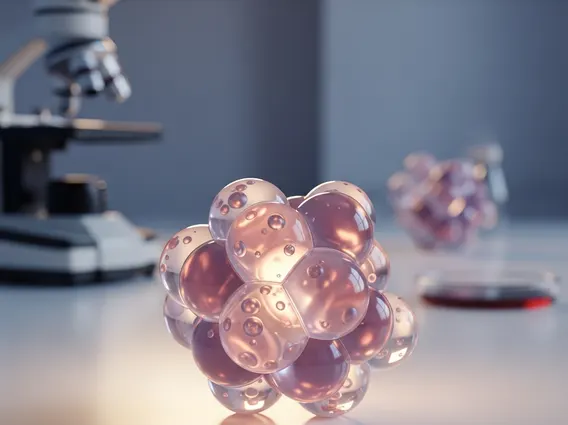
A Hydatidiform Mole is a type of gestational trophoblastic disease (GTD) characterized by the abnormal proliferation of trophoblasts, the cells that normally form the placenta. Instead of developing into a healthy fetus, the placental tissue grows into a mass of fluid-filled cysts resembling a cluster of grapes. This condition is not a viable pregnancy and requires medical intervention.
There are two main types of hydatidiform moles: complete and partial. A complete hydatidiform mole occurs when an egg with no genetic material is fertilized by one or two sperm, leading to only placental tissue growth without any fetal parts. A partial hydatidiform mole occurs when a normal egg is fertilized by two sperm, resulting in an abnormal embryo that is typically non-viable, alongside abnormal placental tissue. According to the American College of Obstetricians and Gynecologists (ACOG), molar pregnancies occur in approximately 1 in 1,000 to 1 in 1,200 pregnancies in the United States.
Symptoms and Causes of Hydatidiform Mole
Recognizing **hydatidiform mole symptoms** is crucial for early diagnosis and management. While some symptoms can mimic those of a normal pregnancy, certain signs are indicative of a molar pregnancy. Common symptoms include:
- Vaginal bleeding, which can range from light spotting to heavy hemorrhage, often dark brown or bright red.
- Severe nausea and vomiting (hyperemesis gravidarum), more pronounced than typical morning sickness.
- Rapid uterine growth, where the uterus measures larger than expected for the gestational age.
- Extremely high levels of human chorionic gonadotropin (hCG), the pregnancy hormone.
- Absence of fetal heart tones or fetal movement.
- Early onset of preeclampsia (high blood pressure and protein in the urine) before 20 weeks of gestation.
The primary **causes of hydatidiform mole** are genetic errors that occur during the fertilization process. In a complete mole, an empty egg is fertilized by one or two sperm, leading to paternal genetic material only, which then duplicates. In a partial mole, a normal egg is fertilized by two sperm, resulting in an embryo with 69 chromosomes (triploidy) instead of the normal 46. Risk factors for developing a hydatidiform mole include advanced maternal age (over 35 or under 20) and a history of previous molar pregnancies.
Hydatidiform Mole Treatment
Effective **hydatidiform mole treatment** primarily involves the removal of the abnormal tissue from the uterus. The most common procedure is dilation and curettage (D&C), where the cervix is dilated, and the molar tissue is gently suctioned or scraped from the uterine lining. In some cases, especially for women who have completed childbearing, a hysterectomy (surgical removal of the uterus) may be considered to prevent recurrence and eliminate the risk of persistent disease.
Following the removal of the molar tissue, meticulous follow-up care is essential. This typically involves weekly monitoring of hCG levels until they return to normal and remain undetectable for several consecutive weeks, usually six months to a year. This monitoring is crucial because, in a small percentage of cases, molar tissue can persist or develop into a more aggressive form of gestational trophoblastic neoplasia (GTN), such as choriocarcinoma. During the follow-up period, contraception is strongly advised to prevent another pregnancy, as a new pregnancy would complicate the interpretation of hCG levels. Any alternative or complementary therapies should be discussed with a healthcare provider, as they are supportive only and do not replace standard medical treatment.