How is Pancreatic Cancer Diagnosed?

How is Pancreatic Cancer Diagnosed?

How is Pancreatic Cancer Diagnosed?

Pancreatic cancer is one of the most challenging malignancies to detect, largely because it rarely causes noticeable symptoms in its earliest stages. Understanding the diagnostic process can help patients and caregivers navigate medical consultations with greater confidence and clarity.

Key Takeaways

  • Pancreatic cancer is frequently diagnosed at an advanced stage due to the absence of early warning symptoms.
  • A combination of imaging scans, blood tests, and tissue biopsy is typically required to confirm the diagnosis.
  • CT scans, MRI, endoscopic ultrasound, and ERCP are among the most commonly used imaging tools.
  • Biopsy remains the definitive method for confirming whether a tumor is malignant.
  • Current research focuses on developing reliable biomarkers and screening protocols for earlier detection.

How is Pancreatic Cancer Diagnosed?

Pancreatic cancer diagnosis methods involve a structured, multi-step clinical evaluation that begins with a thorough review of the patient’s medical history, symptom profile, and physical examination. Because the pancreas lies deep within the abdomen, tumors growing on or within it are not palpable through routine physical exams until they reach a significant size. Physicians typically order a series of tests when symptoms such as jaundice, unexplained weight loss, abdominal pain, or new-onset diabetes raise clinical suspicion.

Blood tests play a supporting role in the diagnostic workup. The tumor marker CA 19-9 is commonly measured, though it is not specific enough to confirm pancreatic cancer on its own. Elevated levels may also appear in benign conditions such as pancreatitis or bile duct obstruction. Liver function tests and complete blood counts help assess overall health and identify signs of biliary obstruction or systemic disease. These laboratory findings guide clinicians toward the appropriate next steps in imaging and tissue sampling.

According to the American Cancer Society, the five-year relative survival rate for pancreatic cancer diagnosed at the localized stage is approximately 44%, compared to just 3% when the disease is detected after distant metastasis. This stark contrast underscores the critical importance of pursuing timely and accurate diagnosis. When a patient presents with relevant risk factors — including a family history of pancreatic cancer, chronic pancreatitis, smoking, obesity, or certain inherited gene mutations — clinicians may adopt a more proactive diagnostic approach.

Imaging Tests and Scans Used to Detect Pancreatic Cancer

Several advanced imaging technologies are integral to identifying pancreatic tumors, assessing their size and location, and determining whether cancer has spread to nearby structures or distant organs. The choice of imaging modality depends on clinical presentation, available resources, and the stage of the diagnostic workup.

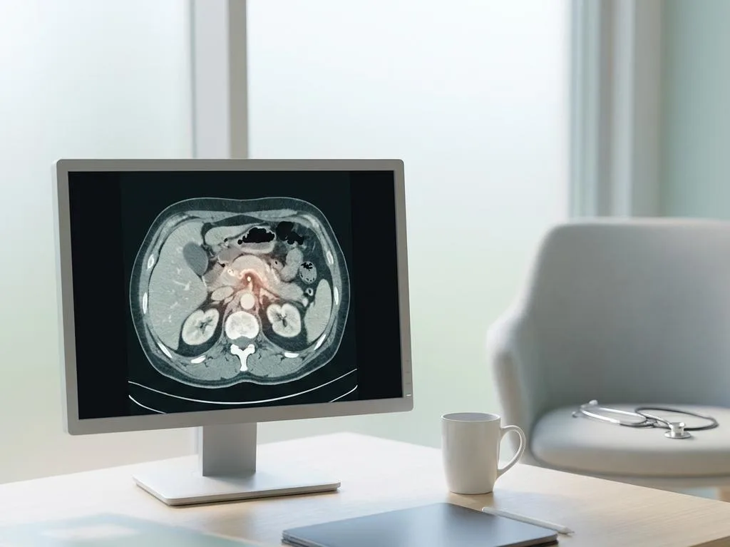
Computed tomography (CT) scanning is generally the first-line imaging test for suspected pancreatic cancer. A multiphasic contrast-enhanced CT scan provides detailed cross-sectional images of the pancreas and surrounding vasculature, helping clinicians assess tumor resectability — that is, whether surgery to remove the tumor is feasible. Magnetic resonance imaging (MRI), often combined with magnetic resonance cholangiopancreatography (MRCP), offers superior soft-tissue contrast and is particularly useful for evaluating the bile and pancreatic ducts without radiation exposure.

Endoscopic ultrasound (EUS) is another valuable tool in pancreatic cancer diagnostic procedures. During EUS, a thin flexible tube with an ultrasound probe is passed through the mouth into the stomach or small intestine, allowing detailed visualization of the pancreas from within the gastrointestinal tract. This proximity produces higher-resolution images than external ultrasound and also permits guided needle biopsy of suspicious lesions. Endoscopic retrograde cholangiopancreatography (ERCP) is used when bile duct obstruction is present, enabling both imaging and therapeutic interventions such as stent placement to relieve jaundice.

Positron emission tomography (PET) scans combined with CT are sometimes employed to detect metastatic spread. A radioactive glucose tracer accumulates in metabolically active cancer cells, producing areas of increased uptake visible on the scan. The following table summarizes the key imaging modalities and their primary diagnostic roles:

Imaging Test Primary Role Key Advantage
CT Scan (contrast-enhanced) Initial tumor detection and staging Fast, widely available, assesses vascular involvement
MRI / MRCP Duct evaluation, soft-tissue detail No radiation; excellent for ductal anatomy
Endoscopic Ultrasound (EUS) High-resolution tumor imaging and biopsy guidance Allows simultaneous tissue sampling
ERCP Bile duct assessment and obstruction relief Combines diagnostic and therapeutic capability
PET-CT Metastasis detection Identifies distant spread not visible on CT alone

Pancreatic Cancer Biopsy: Confirming the Diagnosis

While imaging studies can strongly suggest the presence of a pancreatic tumor, a tissue biopsy remains the definitive method for confirming a malignant diagnosis. Obtaining a cellular sample allows pathologists to examine the tissue under a microscope and determine whether cancer cells are present, as well as classify the type and grade of the tumor. This information is essential for planning appropriate treatment.

Pancreatic cancer biopsy and imaging tests are complementary components of the diagnostic process. The most common biopsy technique is endoscopic ultrasound-guided fine needle aspiration (EUS-FNA), in which a thin needle is passed through the endoscope and directed into the suspicious tissue under real-time ultrasound guidance. This approach is minimally invasive, carries a low complication rate, and yields sufficient cellular material for pathological analysis in the majority of cases. Fine needle biopsy (FNB) using core needles is an alternative that provides slightly larger tissue samples, potentially improving diagnostic accuracy.

In cases where the tumor is not accessible via endoscopic routes — for example, when it has spread to the liver or peritoneum — a CT-guided percutaneous needle biopsy may be performed. A radiologist uses CT imaging to guide a needle through the skin and into the target lesion. For patients who are surgical candidates and whose imaging findings are strongly consistent with resectable pancreatic cancer, some institutions proceed directly to surgery without a preoperative biopsy, as the biopsy result would not alter the treatment decision. Molecular and genetic testing of the biopsy specimen is increasingly performed to identify actionable mutations that may influence systemic therapy selection.

Early Detection of Pancreatic Cancer: Challenges and Current Approaches

Early detection of pancreatic cancer presents a formidable clinical challenge. The pancreas is a retroperitoneal organ, meaning it sits behind the stomach and is largely shielded from routine examination. Tumors can grow for months or even years before causing symptoms significant enough to prompt medical evaluation. As a result, more than half of all cases are diagnosed only after the cancer has metastasized, according to data from the National Cancer Institute’s Surveillance, Epidemiology, and End Results (SEER) program.

Unlike cancers such as breast, cervical, or colorectal cancer, pancreatic cancer currently lacks an established, evidence-based population-wide screening program. For average-risk individuals, no standard screening test has demonstrated sufficient sensitivity and specificity to justify routine use. However, high-risk individuals — including those with certain hereditary syndromes (such as BRCA2 mutations, Lynch syndrome, or familial atypical multiple mole melanoma syndrome) and those with a strong family history of pancreatic cancer — are candidates for structured surveillance. Major cancer centers recommend annual EUS or MRI/MRCP screening for this population, beginning at age 50 or ten years before the youngest affected relative’s diagnosis age.

Research into blood-based biomarkers and liquid biopsy technology represents one of the most promising avenues for improving early detection. Liquid biopsy involves analyzing circulating tumor DNA (ctDNA), circulating tumor cells, or exosomal proteins in a patient’s blood sample without requiring tissue extraction. Several investigational multi-cancer early detection (MCED) tests are currently under clinical evaluation and may eventually offer a non-invasive way to identify pancreatic cancer at an earlier, more treatable stage. Until validated tools become broadly available, maintaining clinical awareness of risk factors and acting promptly on suspicious symptoms remain the most practical strategies for earlier diagnosis.

Frequently Asked Questions

Can a blood test alone confirm pancreatic cancer?

No blood test alone can confirm pancreatic cancer. The CA 19-9 tumor marker is frequently measured but lacks the sensitivity and specificity needed for a definitive diagnosis. Elevated CA 19-9 can also occur in benign conditions such as pancreatitis or bile duct obstruction. Blood tests are used alongside imaging and biopsy as part of a broader diagnostic workup, rather than as standalone confirmation tools.

Is pancreatic cancer visible on a standard abdominal ultrasound?

Standard transabdominal ultrasound can sometimes detect larger pancreatic tumors or signs of bile duct dilation, but it is generally not reliable for identifying smaller lesions due to the pancreas’s deep location and interference from bowel gas. When pancreatic cancer is suspected, physicians typically proceed to more advanced imaging such as contrast-enhanced CT, MRI, or endoscopic ultrasound for a clearer and more detailed assessment.

Who should be screened for pancreatic cancer?

Routine population-wide screening is not currently recommended. Surveillance is advised for high-risk individuals, including those with hereditary syndromes linked to increased pancreatic cancer risk, those with two or more first-degree relatives affected by the disease, and carriers of gene mutations such as BRCA1, BRCA2, PALB2, or ATM. These individuals are typically monitored with annual EUS or MRI starting at age 50 or earlier, depending on family history.


[EN] Cancer Types
Cancer Clinical Trial Options

Specialized matching specifically for oncology clinical trials and cancer care research.

Your Birthday


By filling out this form, you're consenting only to release your medical records. You're not agreeing to participate in clinical trials yet.

Pancreatic cancer is one of the most challenging malignancies to detect, largely because it rarely causes noticeable symptoms in its earliest stages. Understanding the diagnostic process can help patients and caregivers navigate medical consultations with greater confidence and clarity.

Key Takeaways

  • Pancreatic cancer is frequently diagnosed at an advanced stage due to the absence of early warning symptoms.
  • A combination of imaging scans, blood tests, and tissue biopsy is typically required to confirm the diagnosis.
  • CT scans, MRI, endoscopic ultrasound, and ERCP are among the most commonly used imaging tools.
  • Biopsy remains the definitive method for confirming whether a tumor is malignant.
  • Current research focuses on developing reliable biomarkers and screening protocols for earlier detection.

How is Pancreatic Cancer Diagnosed?

Pancreatic cancer diagnosis methods involve a structured, multi-step clinical evaluation that begins with a thorough review of the patient’s medical history, symptom profile, and physical examination. Because the pancreas lies deep within the abdomen, tumors growing on or within it are not palpable through routine physical exams until they reach a significant size. Physicians typically order a series of tests when symptoms such as jaundice, unexplained weight loss, abdominal pain, or new-onset diabetes raise clinical suspicion.

Blood tests play a supporting role in the diagnostic workup. The tumor marker CA 19-9 is commonly measured, though it is not specific enough to confirm pancreatic cancer on its own. Elevated levels may also appear in benign conditions such as pancreatitis or bile duct obstruction. Liver function tests and complete blood counts help assess overall health and identify signs of biliary obstruction or systemic disease. These laboratory findings guide clinicians toward the appropriate next steps in imaging and tissue sampling.

According to the American Cancer Society, the five-year relative survival rate for pancreatic cancer diagnosed at the localized stage is approximately 44%, compared to just 3% when the disease is detected after distant metastasis. This stark contrast underscores the critical importance of pursuing timely and accurate diagnosis. When a patient presents with relevant risk factors — including a family history of pancreatic cancer, chronic pancreatitis, smoking, obesity, or certain inherited gene mutations — clinicians may adopt a more proactive diagnostic approach.

Imaging Tests and Scans Used to Detect Pancreatic Cancer

Several advanced imaging technologies are integral to identifying pancreatic tumors, assessing their size and location, and determining whether cancer has spread to nearby structures or distant organs. The choice of imaging modality depends on clinical presentation, available resources, and the stage of the diagnostic workup.

Computed tomography (CT) scanning is generally the first-line imaging test for suspected pancreatic cancer. A multiphasic contrast-enhanced CT scan provides detailed cross-sectional images of the pancreas and surrounding vasculature, helping clinicians assess tumor resectability — that is, whether surgery to remove the tumor is feasible. Magnetic resonance imaging (MRI), often combined with magnetic resonance cholangiopancreatography (MRCP), offers superior soft-tissue contrast and is particularly useful for evaluating the bile and pancreatic ducts without radiation exposure.

Endoscopic ultrasound (EUS) is another valuable tool in pancreatic cancer diagnostic procedures. During EUS, a thin flexible tube with an ultrasound probe is passed through the mouth into the stomach or small intestine, allowing detailed visualization of the pancreas from within the gastrointestinal tract. This proximity produces higher-resolution images than external ultrasound and also permits guided needle biopsy of suspicious lesions. Endoscopic retrograde cholangiopancreatography (ERCP) is used when bile duct obstruction is present, enabling both imaging and therapeutic interventions such as stent placement to relieve jaundice.

Positron emission tomography (PET) scans combined with CT are sometimes employed to detect metastatic spread. A radioactive glucose tracer accumulates in metabolically active cancer cells, producing areas of increased uptake visible on the scan. The following table summarizes the key imaging modalities and their primary diagnostic roles:

Imaging Test Primary Role Key Advantage
CT Scan (contrast-enhanced) Initial tumor detection and staging Fast, widely available, assesses vascular involvement
MRI / MRCP Duct evaluation, soft-tissue detail No radiation; excellent for ductal anatomy
Endoscopic Ultrasound (EUS) High-resolution tumor imaging and biopsy guidance Allows simultaneous tissue sampling
ERCP Bile duct assessment and obstruction relief Combines diagnostic and therapeutic capability
PET-CT Metastasis detection Identifies distant spread not visible on CT alone

Pancreatic Cancer Biopsy: Confirming the Diagnosis

While imaging studies can strongly suggest the presence of a pancreatic tumor, a tissue biopsy remains the definitive method for confirming a malignant diagnosis. Obtaining a cellular sample allows pathologists to examine the tissue under a microscope and determine whether cancer cells are present, as well as classify the type and grade of the tumor. This information is essential for planning appropriate treatment.

Pancreatic cancer biopsy and imaging tests are complementary components of the diagnostic process. The most common biopsy technique is endoscopic ultrasound-guided fine needle aspiration (EUS-FNA), in which a thin needle is passed through the endoscope and directed into the suspicious tissue under real-time ultrasound guidance. This approach is minimally invasive, carries a low complication rate, and yields sufficient cellular material for pathological analysis in the majority of cases. Fine needle biopsy (FNB) using core needles is an alternative that provides slightly larger tissue samples, potentially improving diagnostic accuracy.

In cases where the tumor is not accessible via endoscopic routes — for example, when it has spread to the liver or peritoneum — a CT-guided percutaneous needle biopsy may be performed. A radiologist uses CT imaging to guide a needle through the skin and into the target lesion. For patients who are surgical candidates and whose imaging findings are strongly consistent with resectable pancreatic cancer, some institutions proceed directly to surgery without a preoperative biopsy, as the biopsy result would not alter the treatment decision. Molecular and genetic testing of the biopsy specimen is increasingly performed to identify actionable mutations that may influence systemic therapy selection.

Early Detection of Pancreatic Cancer: Challenges and Current Approaches

Early detection of pancreatic cancer presents a formidable clinical challenge. The pancreas is a retroperitoneal organ, meaning it sits behind the stomach and is largely shielded from routine examination. Tumors can grow for months or even years before causing symptoms significant enough to prompt medical evaluation. As a result, more than half of all cases are diagnosed only after the cancer has metastasized, according to data from the National Cancer Institute’s Surveillance, Epidemiology, and End Results (SEER) program.

Unlike cancers such as breast, cervical, or colorectal cancer, pancreatic cancer currently lacks an established, evidence-based population-wide screening program. For average-risk individuals, no standard screening test has demonstrated sufficient sensitivity and specificity to justify routine use. However, high-risk individuals — including those with certain hereditary syndromes (such as BRCA2 mutations, Lynch syndrome, or familial atypical multiple mole melanoma syndrome) and those with a strong family history of pancreatic cancer — are candidates for structured surveillance. Major cancer centers recommend annual EUS or MRI/MRCP screening for this population, beginning at age 50 or ten years before the youngest affected relative’s diagnosis age.

Research into blood-based biomarkers and liquid biopsy technology represents one of the most promising avenues for improving early detection. Liquid biopsy involves analyzing circulating tumor DNA (ctDNA), circulating tumor cells, or exosomal proteins in a patient’s blood sample without requiring tissue extraction. Several investigational multi-cancer early detection (MCED) tests are currently under clinical evaluation and may eventually offer a non-invasive way to identify pancreatic cancer at an earlier, more treatable stage. Until validated tools become broadly available, maintaining clinical awareness of risk factors and acting promptly on suspicious symptoms remain the most practical strategies for earlier diagnosis.

Frequently Asked Questions

Can a blood test alone confirm pancreatic cancer?

No blood test alone can confirm pancreatic cancer. The CA 19-9 tumor marker is frequently measured but lacks the sensitivity and specificity needed for a definitive diagnosis. Elevated CA 19-9 can also occur in benign conditions such as pancreatitis or bile duct obstruction. Blood tests are used alongside imaging and biopsy as part of a broader diagnostic workup, rather than as standalone confirmation tools.

Is pancreatic cancer visible on a standard abdominal ultrasound?

Standard transabdominal ultrasound can sometimes detect larger pancreatic tumors or signs of bile duct dilation, but it is generally not reliable for identifying smaller lesions due to the pancreas’s deep location and interference from bowel gas. When pancreatic cancer is suspected, physicians typically proceed to more advanced imaging such as contrast-enhanced CT, MRI, or endoscopic ultrasound for a clearer and more detailed assessment.

Who should be screened for pancreatic cancer?

Routine population-wide screening is not currently recommended. Surveillance is advised for high-risk individuals, including those with hereditary syndromes linked to increased pancreatic cancer risk, those with two or more first-degree relatives affected by the disease, and carriers of gene mutations such as BRCA1, BRCA2, PALB2, or ATM. These individuals are typically monitored with annual EUS or MRI starting at age 50 or earlier, depending on family history.

[EN] Cancer Types
Cancer Clinical Trial Options

Specialized matching specifically for oncology clinical trials and cancer care research.

Your Birthday


By filling out this form, you're consenting only to release your medical records. You're not agreeing to participate in clinical trials yet.

Massive Bio has onboarded over 160,000+ cancer patients to find their clinical trial

Most Recent Article