Hodgkin And Reed Sternberg Cell
Hodgkin lymphoma is a type of cancer that originates in white blood cells called lymphocytes. A hallmark of this disease is the presence of a specific, abnormal cell known as the Hodgkin And Reed Sternberg Cell, which plays a critical role in its diagnosis and progression.
Key Takeaways
- Reed-Sternberg cells are large, abnormal lymphocytes characteristic of Hodgkin lymphoma.
- Their distinctive morphology, often described as “owl’s eye” appearance, is crucial for diagnosis.
- These cells are derived from B-lymphocytes but have lost many typical B-cell features.
- While rare within the tumor mass, their presence is essential for classifying classical Hodgkin lymphoma.
- Understanding these cells is vital for diagnosing and developing targeted treatments for Hodgkin lymphoma.
What is Hodgkin And Reed Sternberg Cell?
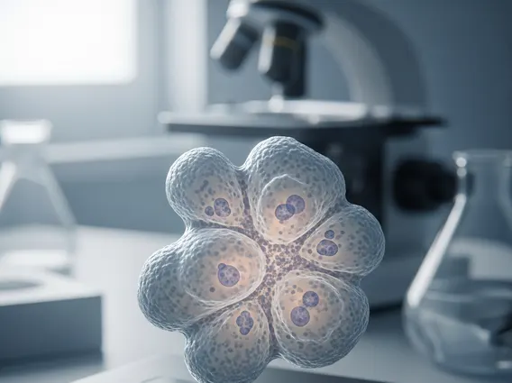
Reed-Sternberg cells are giant, multinucleated cells that are pathognomonic for classical Hodgkin lymphoma. These cells are typically derived from B-lymphocytes, although they have undergone significant genetic and morphological changes, often losing most of their characteristic B-cell markers. Their presence is a definitive diagnostic criterion for classical Hodgkin lymphoma, distinguishing it from other lymphomas and conditions. While they are the defining feature, they often constitute a small percentage of the total cells within the tumor microenvironment, which is predominantly composed of reactive inflammatory cells.
The identification of these cells is pivotal in the diagnostic process. Pathologists examine tissue biopsies, typically from lymph nodes, under a microscope to identify these distinctive cells. The surrounding inflammatory infiltrate, which includes lymphocytes, plasma cells, eosinophils, and histiocytes, is often a reaction to cytokines secreted by the Reed-Sternberg cells themselves, contributing to the overall tumor mass.
Key Characteristics of Reed-Sternberg Cells
The unique morphology of Reed-Sternberg cells is central to their identification and understanding their role in the disease. When considering reed sternberg cell characteristics hodgkin, several features stand out under microscopic examination:
- Size: They are exceptionally large, often 30–100 micrometers in diameter, significantly larger than normal lymphocytes.
- Nuclei: They typically possess two or more nuclei, or a single bilobed nucleus with prominent, eosinophilic nucleoli, often giving them a characteristic “owl’s eye” appearance.
- Cytoplasm: The cytoplasm is abundant and amphophilic (staining with both acidic and basic dyes).
- Immunophenotype: They usually express CD30 and CD15 surface markers, while generally lacking common leukocyte antigen (CD45) and most B-cell specific markers (like CD20), which helps differentiate them from other lymphomas.
- Origin: Despite their altered appearance, genetic studies confirm their origin from germinal center B-cells in most cases of classical Hodgkin lymphoma.
These distinct features allow pathologists to accurately identify and classify Hodgkin lymphoma, guiding subsequent treatment decisions. Variant forms, such as Hodgkin cells (mononuclear Reed-Sternberg cell variants) and lacunar cells (found in nodular sclerosis Hodgkin lymphoma), also contribute to the spectrum of cell types observed.
Role in Hodgkin Lymphoma Development
The hodgkin lymphoma reed sternberg cell function is not fully understood, but it is clear they are the malignant cells that drive the disease. These cells secrete various cytokines and chemokines that recruit and activate the surrounding inflammatory cells, creating a supportive microenvironment. This microenvironment not only protects the Reed-Sternberg cells from the immune system but also provides growth factors that promote their survival and proliferation. This complex interplay between the malignant cells and the reactive infiltrate is a defining feature of Hodgkin lymphoma.
Understanding hodgkin lymphoma cell types explained is crucial for both diagnosis and prognosis. While Reed-Sternberg cells are the malignant component, the bulk of the tumor mass in classical Hodgkin lymphoma consists of non-malignant inflammatory cells. According to the World Health Organization (WHO), Hodgkin lymphoma accounts for about 0.5% of all new cancer cases globally, with an estimated 80,000 new cases diagnosed each year. The presence and characteristics of Reed-Sternberg cells are fundamental to the diagnosis and subtyping of this disease, which significantly impacts treatment strategies and patient outcomes. Modern therapies often target not only the Reed-Sternberg cells but also elements of their supportive microenvironment, leading to improved prognosis for many patients.