Histiocytic Neoplasm
Histiocytic Neoplasm refers to a rare and diverse group of disorders characterized by the abnormal proliferation of histiocytes, which are a type of immune cell. These conditions can affect various organs and tissues throughout the body, leading to a wide range of clinical manifestations.

Key Takeaways
- Histiocytic Neoplasm is a rare group of diseases involving the abnormal growth of histiocytes, a type of immune cell.
- These neoplasms are broadly categorized into Langerhans cell and non-Langerhans cell histiocytoses, each with distinct subtypes.
- While the exact causes are often unknown, genetic mutations play a significant role in their development.
- Symptoms vary greatly depending on the affected organs, ranging from skin lesions and bone pain to organ dysfunction.
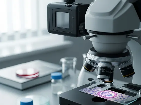
- Diagnosis typically involves biopsy and advanced imaging, with treatment tailored to the specific type and extent of the disease.
What is Histiocytic Neoplasm?
Histiocytic Neoplasm is a collective term for a group of uncommon diseases that arise from the uncontrolled growth of histiocytes. Histiocytes are specialized immune cells, including macrophages, dendritic cells, and Langerhans cells, which play crucial roles in the body’s immune response by engulfing foreign substances and presenting antigens. In these neoplastic conditions, these cells multiply excessively and can infiltrate various tissues, leading to inflammation, tissue damage, and organ dysfunction. These disorders are relatively rare, with an estimated incidence of less than 1 in 100,000 people per year, according to data from various cancer registries.
The clinical presentation of Histiocytic Neoplasm can be highly variable, ranging from localized lesions to widespread systemic disease. The impact on a patient’s health depends heavily on the specific type of histiocyte involved, the organs affected, and the aggressiveness of the cellular proliferation. Early and accurate diagnosis is critical for effective management due to the diverse nature of these conditions.
Types and Causes of Histiocytic Neoplasm
The classification of types of histiocytic neoplasm is complex, but they are generally divided into two main categories: Langerhans cell histiocytosis (LCH) and non-Langerhans cell histiocytosis. LCH involves the proliferation of Langerhans cells, which are typically found in the skin and mucous membranes, but can affect bones, lungs, liver, and other organs. Non-Langerhans cell histiocytoses encompass a broader spectrum of disorders involving other types of histiocytes.
Some prominent types include:
- Langerhans Cell Histiocytosis (LCH): The most common type, affecting children and adults, often presenting with bone lesions, skin rashes, or lung involvement.
- Erdheim-Chester Disease (ECD): A rare, non-Langerhans cell histiocytosis characterized by the infiltration of foamy histiocytes into various tissues, commonly affecting long bones, the cardiovascular system, and the central nervous system.
- Rosai-Dorfman Disease (RDD): Also known as sinus histiocytosis with massive lymphadenopathy, it typically causes painless enlargement of lymph nodes, but can also affect extranodal sites like skin, bone, and the central nervous system.
- Juvenile Xanthogranuloma (JXG): Primarily affects infants and young children, often presenting as benign skin nodules, though systemic forms can occur.
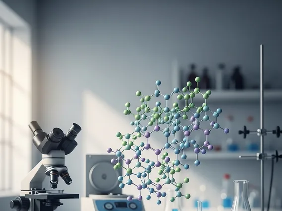
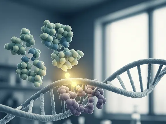
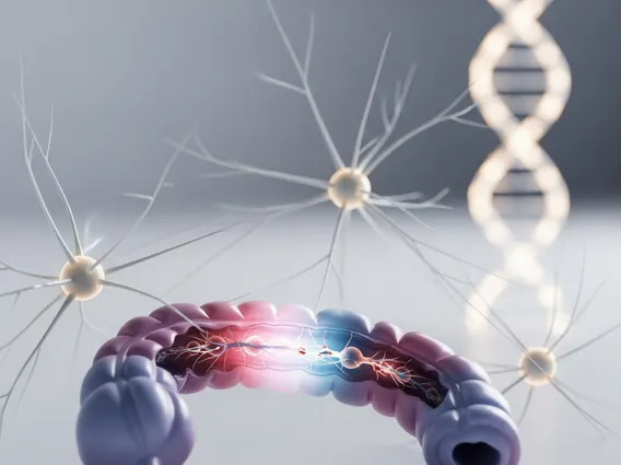
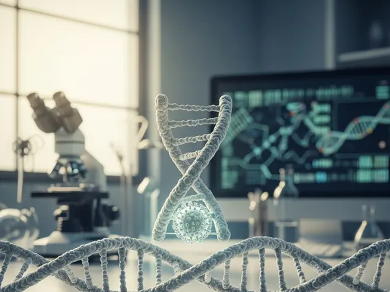
The exact histiocytic neoplasm causes are largely unknown, but research indicates that genetic mutations play a significant role in their development. For instance, mutations in the BRAF gene are frequently found in LCH and ECD, leading to uncontrolled cell growth. Other genetic alterations, such as those in the MAP2K1 and NRAS genes, have also been identified in various histiocytic disorders. These mutations often activate signaling pathways that promote cell proliferation and survival. Environmental factors are not clearly established as primary causes, and these conditions are generally not considered hereditary in the typical sense, though genetic predispositions might exist in some cases.
Symptoms of Histiocytic Neoplasm
The histiocytic neoplasm symptoms are highly variable and depend on the specific type of neoplasm, the organs involved, and the extent of the disease. Because histiocytes can infiltrate nearly any tissue in the body, symptoms can range from mild and localized to severe and life-threatening. Early symptoms are often non-specific, making diagnosis challenging.
Common symptoms and affected areas may include:
| Body System | Potential Symptoms |
|---|---|
| Skin | Rashes (often scaly or reddish-brown), nodules, papules, or yellow-orange lesions. |
| Bones | Localized pain, swelling, tenderness, pathological fractures, or lytic lesions visible on imaging. |
| Lymph Nodes | Painless enlargement, particularly in the neck, armpits, or groin. |
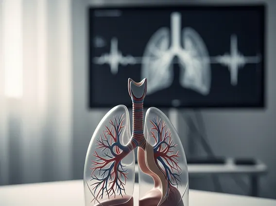
| Lungs | Chronic cough, shortness of breath, chest pain, or recurrent lung infections. |
| Liver/Spleen | Enlargement (hepatomegaly/splenomegaly), abdominal pain, jaundice, or impaired organ function. |
| Endocrine System | Diabetes insipidus (excessive thirst and urination), growth delays, or hormonal imbalances. |
| Central Nervous System | Headaches, seizures, balance problems, vision changes, or neurological deficits. |
| Systemic | Fever, fatigue, weight loss, night sweats, or general malaise. |
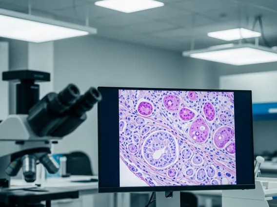
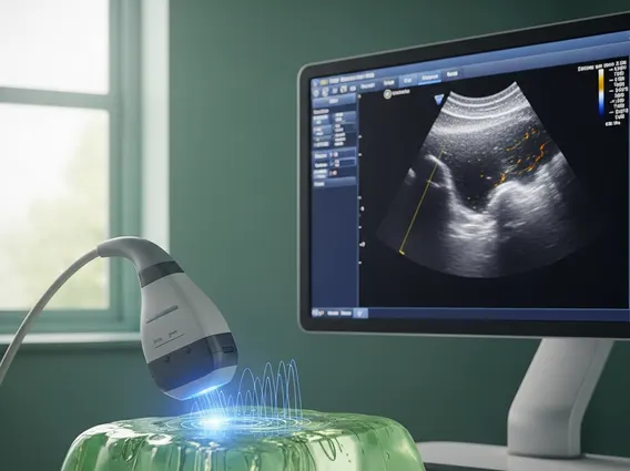
Due to the broad spectrum of possible symptoms, a comprehensive diagnostic approach involving biopsies, imaging studies (such as X-rays, CT scans, and MRI), and genetic testing is often necessary to confirm a diagnosis and determine the extent of the disease. Prompt recognition of these varied symptoms is crucial for timely intervention and management of Histiocytic Neoplasm.
The information provided in this article is for general knowledge and informational purposes only, and does not constitute medical advice. It is essential to consult with a qualified healthcare professional for any health concerns or before making any decisions related to your health or treatment.