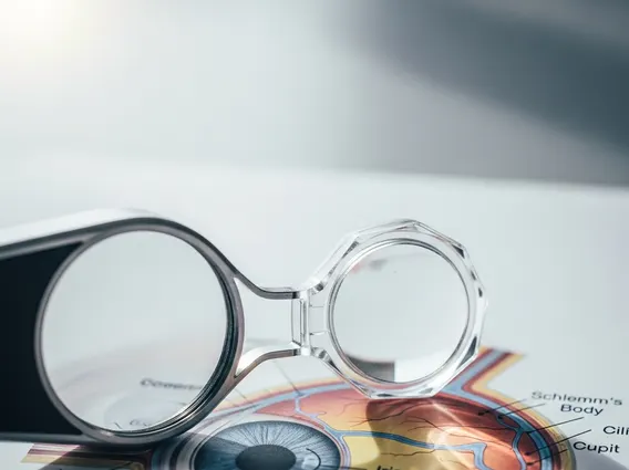
Gonioscopy
Gonioscopy is a specialized eye examination used to visualize the anterior chamber angle of the eye. This crucial diagnostic tool helps ophthalmologists assess the drainage system of the eye, which is vital for maintaining healthy intraocular pressure.
Key Takeaways
- Gonioscopy is an essential eye examination that allows for direct visualization of the eye’s anterior chamber angle.
- It helps evaluate the intricate drainage system of the eye, which is critical for regulating intraocular pressure.
- The procedure involves the use of a special mirrored lens and is generally quick, non-invasive, and well-tolerated.
- It is indispensable for the accurate diagnosis, classification, and ongoing management of various eye conditions, particularly glaucoma.
What is Gonioscopy?
Gonioscopy is a clinical procedure performed by an eye care professional to examine the anterior chamber angle, which is the area inside the eye where the iris meets the cornea. This angle houses the trabecular meshwork, a sponge-like tissue responsible for draining aqueous humor, the fluid that nourishes the eye and maintains its shape. A healthy anterior chamber angle ensures proper fluid outflow, which in turn regulates intraocular pressure (IOP).
During a gonioscopy exam, the ophthalmologist uses a special contact lens, called a gonioscope, in conjunction with a slit lamp microscope. This lens contains mirrors that allow the doctor to see the angle structures that would otherwise be obscured by the limbus (the junction of the cornea and sclera). The ability to directly visualize these structures is fundamental for identifying abnormalities that could impede fluid drainage and lead to elevated IOP.
The Gonioscopy Procedure Explained
The gonioscopy procedure explained involves a straightforward and typically painless process. Patients are usually seated comfortably at a slit lamp microscope, similar to a routine eye exam. Numbing eye drops are applied to ensure comfort and prevent blinking, as the gonioscope lens will gently touch the surface of the eye.
The steps involved in a typical gonioscopy examination include:
- Preparation: The patient receives anesthetic eye drops to numb the eye surface. Pupil dilation is generally not required for gonioscopy.
- Positioning: The patient rests their chin on a chin rest and their forehead against a headrest on the slit lamp.
- Lens Application: The ophthalmologist gently places the gonioscope lens directly onto the surface of the anesthetized eye. This lens has several mirrors angled to reflect light into the anterior chamber angle.
- Examination: The doctor uses the slit lamp to illuminate the eye and systematically view different sections of the angle by rotating the gonioscope lens. They observe key structures like the iris root, ciliary body band, scleral spur, trabecular meshwork, and Schwalbe’s line.
- Completion: Once the examination is complete, the lens is removed. Vision may be slightly blurry for a short period due to the numbing drops or gel used with the lens, but this quickly resolves.
The entire procedure typically takes only a few minutes per eye. Patients can usually resume their normal activities immediately afterward, though they should avoid rubbing their eyes until the numbing effect wears off.
Purpose of Gonioscopy: Diagnosing Glaucoma and Other Conditions
The primary purpose of gonioscopy eye exam is to assess the anterior chamber angle, which is crucial for diagnosing and managing a variety of ocular conditions. It is particularly indispensable for gonioscopy for glaucoma diagnosis. Glaucoma is a group of eye conditions that damage the optic nerve, often caused by abnormally high pressure in the eye. According to the World Health Organization (WHO), glaucoma is the second leading cause of blindness globally, affecting an estimated 64 million people worldwide.
Gonioscopy helps classify glaucoma into its main types: open-angle glaucoma and angle-closure glaucoma. In open-angle glaucoma, the drainage angle appears open but the trabecular meshwork is not functioning efficiently. In angle-closure glaucoma, the iris blocks or narrows the drainage angle, preventing fluid outflow. Identifying the type of glaucoma is critical for determining the most effective treatment strategy, whether it involves medication, laser therapy, or surgery.
Beyond glaucoma, gonioscopy is also vital for detecting and evaluating other conditions, including:
- Ocular Trauma: Identifying blood (hyphema), foreign bodies, or structural damage within the angle.
- Inflammation: Detecting inflammatory cells or deposits (synechiae) that can obstruct the drainage system.
- Tumors: Visualizing abnormal growths or cysts in the angle area.
- Developmental Anomalies: Identifying congenital malformations of the angle structures that may predispose individuals to glaucoma or other issues.
By providing a clear view of the anterior chamber angle, gonioscopy enables ophthalmologists to make accurate diagnoses, monitor disease progression, and tailor treatment plans, thereby playing a pivotal role in preserving vision and maintaining overall eye health.