Gallium Scan
A Gallium Scan is a specialized nuclear medicine imaging test used to detect inflammation, infection, and certain types of tumors within the body. It involves the injection of a small amount of radioactive gallium, which accumulates in areas of increased cellular activity, providing valuable diagnostic information.
Key Takeaways
- A Gallium Scan is a nuclear medicine test that uses a radioactive tracer to identify areas of inflammation, infection, or tumor growth.
- The scan is particularly useful for diagnosing certain infections, inflammatory conditions, and lymphomas.
- The procedure involves an injection of gallium-67, followed by imaging sessions typically 24 to 72 hours later.
- Preparation may include dietary restrictions and bowel cleansing to ensure clear images.
- Results are interpreted by a nuclear medicine physician, looking for areas of increased gallium uptake, which indicate abnormal activity.
What is a Gallium Scan and What is it Used For?
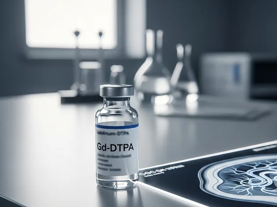
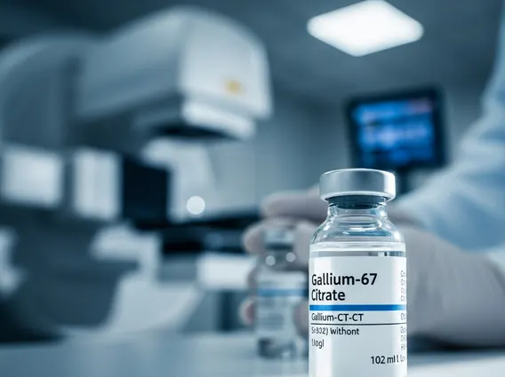
A Gallium Scan is a diagnostic imaging technique that falls under nuclear medicine. It involves the use of a radioactive isotope, typically Gallium-67 citrate, which is injected into the patient’s bloodstream. This radiotracer has a natural affinity for areas of inflammation, infection, and certain types of rapidly dividing cells, such as those found in some tumors. The primary purpose of a Gallium Scan is to pinpoint the location and extent of these abnormal processes within the body.
The applications for what is a gallium scan used for are diverse, making it a valuable tool in specific clinical scenarios. It is frequently employed to:
- Detect and localize occult infections, particularly in cases of fever of unknown origin or suspected osteomyelitis (bone infection).
- Identify inflammatory conditions, such as sarcoidosis, pulmonary fibrosis, or certain autoimmune diseases.
- Stage and monitor certain cancers, most notably lymphomas (e.g., Hodgkin lymphoma and some non-Hodgkin lymphomas), as gallium can accumulate in these malignant cells.
- Assess the activity of chronic infections, like tuberculosis or fungal infections.
While other imaging modalities exist, the Gallium Scan offers unique insights into the metabolic activity of tissues, complementing anatomical imaging techniques like CT or MRI. According to data from the Society of Nuclear Medicine and Molecular Imaging, nuclear medicine procedures like the Gallium Scan contribute significantly to the diagnosis and management of various diseases.
Gallium Scan Procedure: How It Works and Preparation
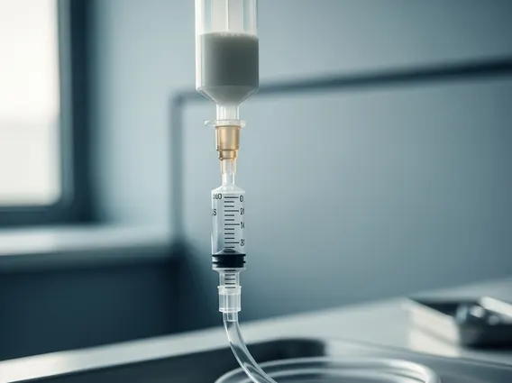
The gallium scan procedure and preparation involves several steps to ensure accurate imaging. The process begins with the intravenous injection of a small, safe dose of Gallium-67 citrate. Once injected, the gallium circulates throughout the body and gradually accumulates in areas of increased cellular activity or inflammation. This accumulation is due to gallium’s chemical similarity to ferric iron, allowing it to bind to iron-binding proteins like transferrin, which are often upregulated in inflammatory and neoplastic tissues.
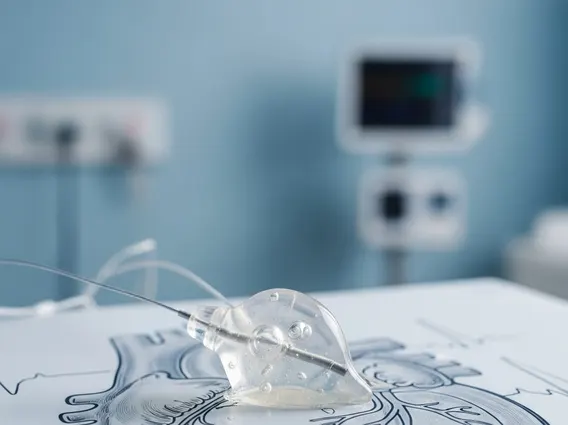
How does a gallium scan work? After the injection, there is a waiting period, typically ranging from 24 to 72 hours, sometimes even up to 5-7 days, to allow the gallium to distribute and concentrate in the target areas while non-target gallium clears from the bloodstream. During this time, patients are generally free to go about their normal activities. Imaging is then performed using a gamma camera, which detects the gamma rays emitted by the Gallium-67. The camera moves slowly over the body, capturing images that are then processed by a computer to create detailed pictures of gallium distribution.
Preparation for a Gallium Scan is crucial for optimal results. Patients may be advised to:
- Inform their doctor about all medications they are taking, as some can interfere with gallium uptake.
- Undergo bowel preparation (e.g., laxatives or enemas) before imaging, especially if abdominal or pelvic pathology is suspected, to reduce background activity from the bowel that could obscure findings.
- Stay well-hydrated to help clear unbound gallium from the body.
- Avoid certain iron supplements, as iron can compete with gallium for binding sites.
The imaging session itself is usually comfortable, requiring the patient to lie still on a table while the camera acquires images. The duration of the scan can vary but typically lasts between 30 minutes to an hour per session.
Interpreting Gallium Scan Results
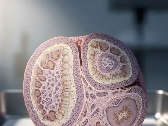
The gallium scan results interpretation is performed by a nuclear medicine physician who specializes in analyzing these types of images. The physician looks for areas where the gallium has accumulated more intensely than in surrounding normal tissues, often referred to as “hot spots.” These hot spots indicate increased metabolic activity, which can be a sign of infection, inflammation, or tumor presence. The intensity and pattern of gallium uptake, along with the location of these hot spots, provide critical clues about the underlying condition.
For instance, diffuse uptake in the lungs might suggest an inflammatory process like sarcoidosis, while focal, intense uptake in a lymph node could point towards lymphoma. In cases of suspected infection, gallium accumulation at a specific site can confirm the presence and location of the infectious process. However, it is important to note that gallium uptake is not specific to a single disease; for example, both infection and tumor can cause increased uptake. Therefore, the results are always interpreted in conjunction with the patient’s clinical history, physical examination, and findings from other diagnostic tests, such as blood work, X-rays, CT scans, or MRI scans.
The physician will generate a detailed report outlining the findings, including the location and intensity of any abnormal gallium uptake. This report is then sent to the referring physician, who will use this information to guide further diagnostic steps, treatment planning, or to monitor the effectiveness of ongoing therapies. While a Gallium Scan is a powerful diagnostic tool, its results are part of a larger clinical picture and are rarely used in isolation for definitive diagnosis.