Galactography
Galactography, also known as ductography, is a specialized diagnostic imaging procedure used to investigate abnormal nipple discharge. This minimally invasive technique helps pinpoint the cause of discharge by visualizing the milk ducts within the breast.

Key Takeaways
- Galactography is a medical imaging procedure specifically designed to evaluate abnormal nipple discharge.
- It involves injecting a contrast dye into a discharging milk duct, followed by mammography, to visualize the ductal system.
- The primary indications include spontaneous, persistent, and bloody or serous nipple discharge from a single duct.
- The procedure helps identify intraductal lesions such as papillomas, fibrocystic changes, or carcinoma, guiding further treatment.
- Despite its name, **Galactography** in the medical context has no relation to the study of galaxies or astronomy.
What is Galactography: Definition and Scope
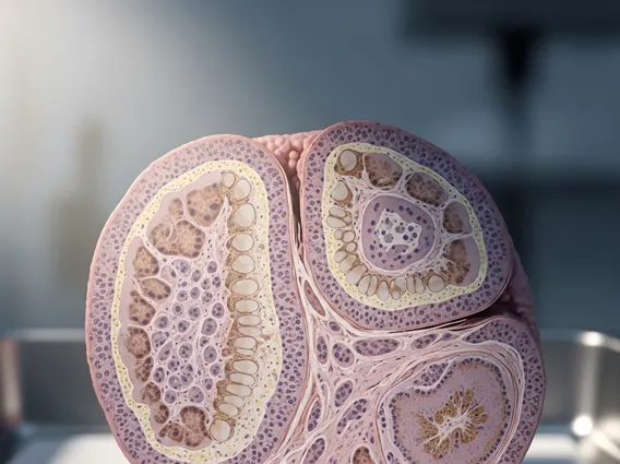
Galactography, often referred to as ductography, is a highly specialized radiological procedure utilized in breast imaging. It involves the careful injection of a small amount of contrast material directly into a milk duct that is actively producing abnormal discharge. Following the injection, mammographic images are taken, which allow radiologists to visualize the internal architecture of the ductal system and identify any abnormalities such as masses, strictures, or dilatations.
The primary purpose of **galactography meaning and scope** is to precisely locate and characterize the source of pathological nipple discharge, which can be a symptom of various benign or malignant conditions. This detailed visualization is crucial for guiding subsequent diagnostic steps or surgical planning. It is important to note that despite the etymological roots of the word, **Galactography** in the medical field has no connection to the “galactography study of galaxies” or the “importance of galactography in astronomy.” This term is exclusively used to describe a diagnostic procedure pertaining to the mammary ducts.
Indications for Galactography
Galactography is typically recommended when a patient experiences spontaneous, persistent, and often unilateral nipple discharge that is not associated with pregnancy or lactation. The characteristics of the discharge are key in determining the need for this procedure. Common indications include:
- Bloody or Serosanguineous Discharge: This type of discharge is a significant concern as it can be associated with intraductal papillomas or, less commonly, ductal carcinoma.
- Serous (Clear or Yellowish) Discharge: While often benign, persistent serous discharge from a single duct warrants investigation.
- Spontaneous and Persistent Discharge: Discharge that occurs without manipulation and continues over time is a primary indication.
- Unilateral and Uniductal Discharge: When the discharge originates from a single pore on one breast, it strongly suggests a localized intraductal pathology.
The procedure helps differentiate between various causes of nipple discharge, ranging from benign conditions like intraductal papillomas or fibrocystic changes to more serious pathologies such as ductal carcinoma in situ (DCIS) or invasive ductal carcinoma. According to the American Cancer Society, nipple discharge accounts for about 5-10% of all breast symptoms, and galactography plays a vital role in evaluating these cases, particularly when clinical examination and conventional mammography are inconclusive.
The Galactography Procedure
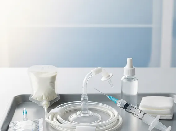
The galactography procedure is performed in an outpatient setting and typically takes about 30 to 60 minutes. The steps involved are meticulous to ensure accurate imaging:
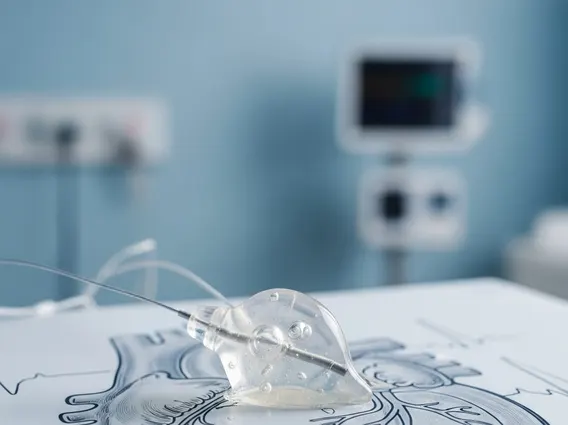
- Preparation: The patient lies comfortably on an examination table. The breast is cleaned, and the specific discharging duct is identified, often by gently compressing the breast to elicit discharge.
- Duct Cannulation: A very fine, blunt-tipped cannula (a thin tube) is carefully inserted into the opening of the discharging milk duct. This step requires precision and a steady hand.
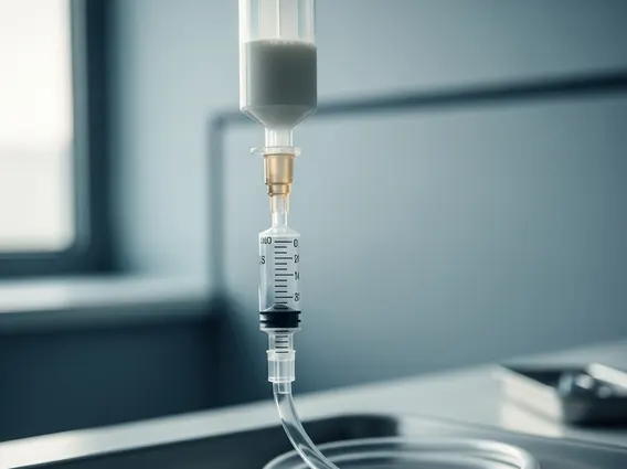
- Contrast Injection: A small amount (typically 0.1 to 0.5 mL) of water-soluble, iodine-based contrast material is slowly injected into the duct. The patient may feel a slight pressure or warmth during this stage.
- Mammography: Immediately after contrast injection, standard mammographic views of the breast are taken. These images capture the contrast material as it fills the ductal system, highlighting any irregularities or lesions within the ducts. Additional views, such as magnification or spot compression, may be performed to obtain clearer details of suspicious areas.
- Post-Procedure Care: After the images are acquired, the cannula is removed. Patients are typically advised to avoid squeezing the nipple for a day or two to prevent further discharge. Mild discomfort or bruising may occur, but serious complications are rare.
The images obtained from galactography are then interpreted by a radiologist to identify the location, size, and characteristics of any intraductal lesions. This information is crucial for the referring physician to determine the most appropriate course of action, which may include surgical excision, further imaging, or clinical observation.