
Endosonography
Endosonography is an advanced medical imaging technique that combines endoscopy with ultrasound technology. This procedure provides highly detailed images of internal organs and structures, particularly those within or adjacent to the gastrointestinal tract and respiratory system.
Key Takeaways
- Endosonography is a minimally invasive procedure combining endoscopy and ultrasound for detailed internal imaging.
- It is crucial for diagnosing and staging various conditions, including cancers of the digestive and respiratory systems.
- The procedure involves inserting a thin, flexible tube with an ultrasound probe through the mouth or rectum.
- Unlike traditional ultrasound, endosonography offers superior resolution of deep tissues by placing the ultrasound transducer closer to the area of interest.
- It aids in precise tissue sampling (biopsy) and guiding therapeutic interventions.
What is Endosonography?
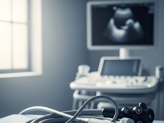
Endosonography, also known as endoscopic ultrasound (EUS), is a sophisticated diagnostic procedure that merges two powerful medical technologies: endoscopy and ultrasound. During an endosonography procedure, a specialized endoscope equipped with a tiny ultrasound transducer at its tip is carefully guided into the body. This allows physicians to obtain high-resolution ultrasound images from inside the gastrointestinal tract or respiratory passages, providing an unparalleled view of the walls of these organs and adjacent structures such as lymph nodes, blood vessels, and surrounding organs like the pancreas, liver, and bile ducts.
This technique offers a unique advantage by positioning the ultrasound probe very close to the area of interest, bypassing obstacles like bone, air, or fat that can obscure images from external ultrasound. The detailed images produced by endosonography are instrumental in the precise diagnosis and staging of various medical conditions, particularly those affecting the digestive and respiratory systems.
Endosonography Procedure and Diagnostic Uses
The endosonography procedure explained involves the patient typically receiving sedation to ensure comfort. A thin, flexible endoscope, fitted with an ultrasound transducer, is then gently advanced through the mouth (for upper GI or respiratory tract) or rectum (for lower GI tract). As the endoscope moves, the ultrasound probe emits sound waves that bounce off internal structures, creating real-time images displayed on a monitor. This allows the physician to visualize layers of the organ wall, surrounding tissues, and nearby lymph nodes with exceptional clarity.
The uses of endosonography diagnosis are extensive and critical for various medical conditions. It is particularly valuable in oncology for detecting, staging, and monitoring cancers within the esophagus, stomach, duodenum, pancreas, rectum, and lungs. Beyond cancer, endosonography is also used to:
- Evaluate abnormalities such as cysts, stones, or strictures in the bile ducts and pancreas.
- Assess chronic pancreatitis and other pancreatic diseases.
- Investigate unexplained abdominal pain or weight loss.
- Determine the depth of penetration of gastrointestinal tumors.
- Guide fine-needle aspiration (FNA) or biopsy of suspicious lesions or lymph nodes, allowing for tissue samples to be collected for pathological analysis.
The ability to perform guided biopsies during the procedure significantly enhances diagnostic accuracy, often eliminating the need for more invasive surgical biopsies.
Endosonography vs. Traditional Ultrasound
Understanding the endosonography vs ultrasound differences is key to appreciating the unique benefits of endosonography. While both techniques use sound waves to create images of internal body structures, their approach and resulting image quality differ significantly. Traditional, or external, ultrasound involves placing a transducer on the skin surface to send and receive sound waves. This method is non-invasive and excellent for broad screening and visualizing superficial organs.
However, traditional ultrasound can be limited by factors such as the patient’s body habitus, overlying bone, or gas in the intestines, which can scatter sound waves and obscure deeper structures. Endosonography overcomes these limitations by placing the ultrasound transducer internally, much closer to the target organs. This proximity allows for the use of higher-frequency sound waves, which provide superior resolution and detailed imaging of the organ walls and adjacent structures that are often inaccessible or poorly visualized with external ultrasound.
The following table highlights key distinctions between the two imaging modalities:
| Feature | Endosonography (EUS) | Traditional Ultrasound |
|---|---|---|
| Transducer Placement | Internal (via endoscope) | External (on skin surface) |
| Image Resolution | High-resolution, detailed images of organ walls and adjacent structures | Lower resolution for deep structures, better for broad screening |
| Penetration Depth | Excellent for deep tissues near GI/respiratory tract | Limited by bone, air, and body habitus for deep structures |
| Invasiveness | Minimally invasive (requires sedation) | Non-invasive |
| Primary Use | Detailed diagnosis, cancer staging, guided biopsies | Initial screening, general organ assessment, obstetrics |
In essence, while traditional ultrasound offers a broad overview, endosonography provides a highly focused, detailed view, making it indispensable for precise diagnosis and intervention in specific clinical scenarios.


