Embryoma
Embryoma refers to a diverse group of tumors that originate from embryonic or fetal tissues. These tumors are typically found in infants and children, though they can occasionally present in adults. They represent a significant area of study in pediatric oncology due to their unique developmental origins and varied clinical presentations.

Key Takeaways
- Embryomas are tumors derived from embryonic cells, predominantly affecting children.
- Symptoms vary widely depending on the tumor’s location and size, often including palpable masses or non-specific signs.
- Diagnosis involves a combination of imaging, biopsy, and pathological examination.
- Treatment typically includes surgery, chemotherapy, and sometimes radiation therapy, tailored to the specific type and stage of the embryoma.
- Early detection and a multidisciplinary approach are crucial for effective management and improved outcomes.
What is Embryoma?
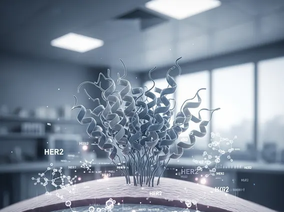
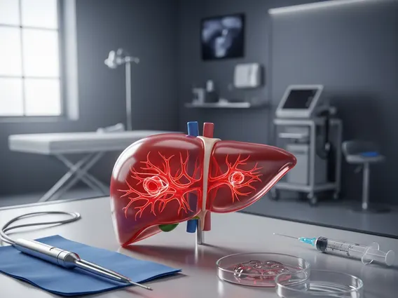
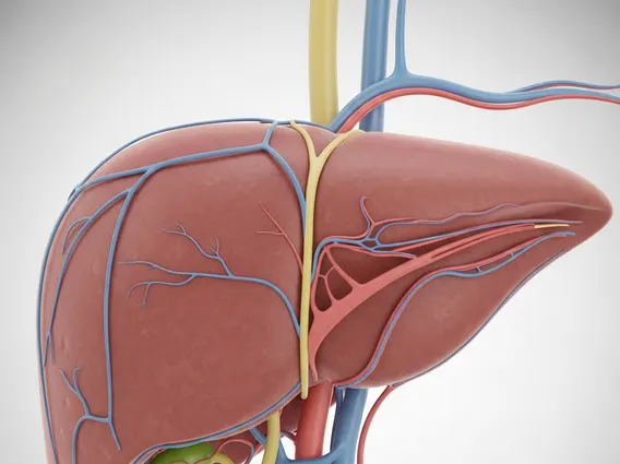
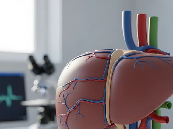
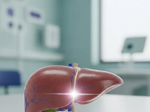
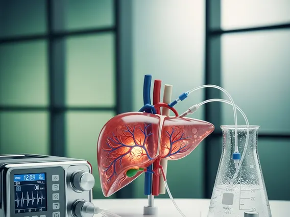
Embryoma is a broad term encompassing neoplasms that arise from primitive, undifferentiated embryonic cells. These tumors are thought to result from errors during fetal development, where certain cells fail to mature properly and instead proliferate uncontrollably. While they are most commonly associated with childhood cancers, their exact nature and behavior can differ significantly based on the specific tissue of origin and the degree of cellular differentiation. Examples include nephroblastoma (Wilms’ tumor) originating in the kidney, neuroblastoma from neural crest cells, and hepatoblastoma from liver precursor cells. These tumors are characterized by their cellular resemblance to embryonic tissues, often containing immature cells that reflect their developmental origins.
Embryoma Symptoms, Causes, and Diagnosis
The presentation of an embryoma is highly variable, largely dependent on its location, size, and the specific organ system affected. Common embryoma symptoms and causes often include a palpable mass in the abdomen, flank, or other body regions. Other non-specific symptoms may include pain, fever, weight loss, fatigue, or changes in bowel or bladder habits. For instance, a kidney embryoma might present with abdominal swelling and hypertension, while a brain embryoma could lead to neurological deficits or seizures. The precise causes are often multifactorial, involving a complex interplay of genetic predispositions and environmental factors, though specific genetic mutations are increasingly identified in various embryoma types.
Diagnosing embryoma typically involves a comprehensive approach. Initial evaluation often includes a thorough physical examination and a detailed medical history. Imaging studies are crucial for identifying the tumor, assessing its size, location, and spread. These may include:
- Ultrasound: Often the first imaging modality, especially for abdominal masses, due to its non-invasive nature.
- Computed Tomography (CT) scans: Provide detailed cross-sectional images, helping to define tumor boundaries and involvement of surrounding structures.
- Magnetic Resonance Imaging (MRI): Offers superior soft tissue contrast, particularly useful for brain, spinal, and complex abdominal embryomas.
- Biopsy: A definitive diagnosis requires a tissue sample obtained through a biopsy, which is then examined by a pathologist. This microscopic analysis confirms the presence of embryonic cells and helps classify the specific type of embryoma, guiding treatment decisions.
- Blood tests: May be used to check for tumor markers, which can aid in diagnosis and monitoring treatment response for certain types of embryomas.
Early and accurate diagnosis is paramount for improving patient outcomes, as it allows for timely initiation of appropriate treatment strategies.
Embryoma Treatment Options
The management of an embryoma is highly individualized, depending on the specific type of tumor, its stage, location, and the patient’s overall health. Embryoma treatment options typically involve a multidisciplinary team of specialists, including pediatric oncologists, surgeons, radiation oncologists, and pathologists. The primary modalities often include surgery, chemotherapy, and radiation therapy.
Surgery is frequently the cornerstone of treatment, aiming for complete removal of the tumor whenever feasible. In some cases, neoadjuvant chemotherapy (chemotherapy given before surgery) may be used to shrink the tumor, making surgical resection easier and safer. Adjuvant chemotherapy (chemotherapy given after surgery) is often administered to eliminate any remaining cancer cells and reduce the risk of recurrence. Radiation therapy may be employed for certain types of embryomas, especially if there is residual disease after surgery or if the tumor has spread to critical areas, such as the brain or lungs. The specific chemotherapy agents, radiation doses, and surgical techniques are carefully chosen based on established protocols for each embryoma subtype, with ongoing research continually refining these approaches to maximize efficacy and minimize side effects.