Electron Microscope
Electron Microscopes are powerful tools that have revolutionized our understanding of biological structures at a nanoscale. In clinical and research settings, they provide unparalleled insights into cellular architecture, pathogen morphology, and disease mechanisms.
Key Takeaways
- Electron Microscopes use electron beams instead of light to visualize ultra-fine details of specimens.
- They offer significantly higher magnification and resolution compared to traditional light microscopes.
- Operating principles involve electron generation, electromagnetic lenses, and signal detection.
- Key types include Transmission Electron Microscopes (TEM) and Scanning Electron Microscopes (SEM).
- Their applications are crucial in medical diagnostics, pathology, virology, and cancer research.
What is an Electron Microscope?
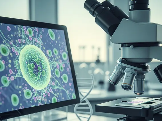
An Electron Microscope is a powerful analytical instrument that employs a beam of accelerated electrons to create highly magnified images of specimens. Unlike traditional optical microscopes which are limited by the wavelength of visible light, electron microscopes utilize electrons, whose much shorter wavelengths allow for significantly greater resolution and magnification, often revealing details at the nanometer scale. This advanced capability makes them indispensable for visualizing the intricate ultrastructure of biological samples, materials, and even individual atoms, providing crucial insights for medical diagnostics, research, and understanding fundamental biological processes.
Operating Principles and Types of Electron Microscopes
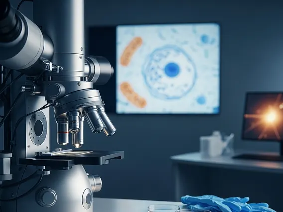
The core principle behind how an electron microscope works involves generating a stream of electrons from an electron gun, which are then accelerated by high voltage and focused onto a specimen using electromagnetic lenses. As these electrons interact with the specimen, they produce various signals that are collected by detectors and processed to form a highly detailed image. This interaction can reveal information about the specimen’s topography, composition, and internal structure, far surpassing the limits of light microscopy.
There are primarily two main types of electron microscopes that are crucial in scientific and clinical settings:
- Transmission Electron Microscope (TEM): A TEM operates by transmitting a beam of electrons through an ultrathin section of a specimen. The electrons that pass through are then focused by objective and projector lenses to form a highly magnified image on a fluorescent screen or digital detector. TEMs provide two-dimensional, high-resolution images of internal structures, making them invaluable for studying cellular organelles, viral particles, and crystalline structures in detail.
- Scanning Electron Microscope (SEM): An SEM, in contrast, scans a finely focused electron beam across the surface of a bulk specimen. The interaction of the electron beam with the specimen’s surface generates various signals, such as secondary electrons and backscattered electrons, which are detected and used to construct a three-dimensional-like image of the specimen’s surface topography and composition. SEMs are widely used for examining surface features of cells, tissues, and materials, offering insights into their morphology and texture.
Applications of Electron Microscopes
The wide-ranging electron microscope applications are fundamental to progress in medical science, particularly in diagnostics and research. In clinical pathology, Electron Microscopes are indispensable for the definitive diagnosis of certain diseases when light microscopy is insufficient. For example, they are critical in identifying specific types of renal diseases, neuromuscular disorders, and storage diseases by revealing characteristic ultrastructural abnormalities in biopsy samples. The ability to visualize cellular pathology at such high resolution allows for precise classification and often guides therapeutic decisions.
In the field of oncology, Electron Microscopes play a vital role in the detailed characterization of tumor cells. They help pathologists differentiate between various tumor types, especially in cases where light microscopic features are ambiguous, such as distinguishing between poorly differentiated carcinomas, lymphomas, and sarcomas. This precise morphological information can be crucial for prognosis and selecting appropriate treatment protocols. Furthermore, in infectious disease research and virology, Electron Microscopes are essential for visualizing the morphology of bacteria and viruses, studying their replication cycles, and understanding their interactions with host cells. This contributes significantly to the development of vaccines and antiviral therapies, making the Electron Microscope an unparalleled tool for advancing medical knowledge and enhancing diagnostic accuracy.