
Cytokeratin
Cytokeratin refers to a family of fibrous structural proteins that are crucial components of the cytoskeleton in epithelial cells. These proteins provide mechanical stability and play a significant role in maintaining cell integrity and tissue architecture.
Key Takeaways
- Cytokeratin proteins form intermediate filaments within epithelial cells, providing structural support and mechanical strength.
- There are over 20 different types of Cytokeratins, classified into Type I (acidic) and Type II (basic/neutral), which typically pair together.
- These proteins are expressed in a tissue-specific manner throughout the human body, contributing to the unique properties of various epithelial tissues.
- Cytokeratin expression patterns are vital biomarkers used in pathology for the diagnosis, classification, and prognosis of various diseases, particularly carcinomas.
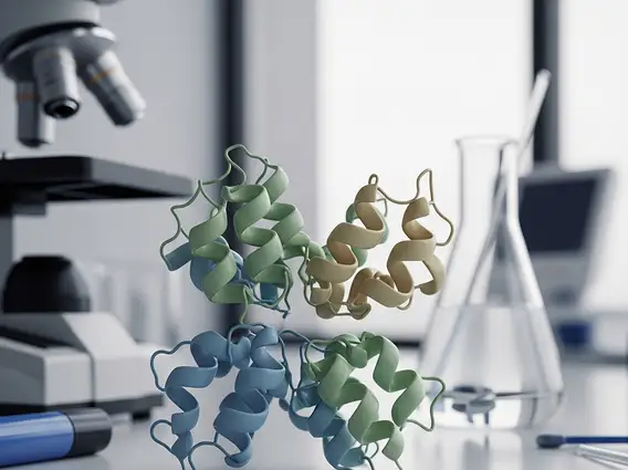
What is Cytokeratin: Structure and Types
Cytokeratin is a class of intermediate filament proteins found exclusively in epithelial cells, which line the surfaces of organs and blood vessels throughout the body. These proteins are essential for the structural integrity and mechanical resilience of epithelial tissues, forming a robust intracellular network that connects to cell junctions and provides resistance against physical stress. The intricate structure of Cytokeratins allows them to withstand significant forces without rupturing, protecting cells from damage.
There are over 20 known types of human Cytokeratins, each with a specific molecular weight and isoelectric point. These types are broadly categorized into two groups: Type I (acidic, K9-K20) and Type II (basic or neutral, K1-K8). For a functional intermediate filament to form, one acidic Cytokeratin must pair with one basic Cytokeratin. The specific combination of these proteins varies depending on the type of epithelial cell and its developmental stage, contributing to the diverse mechanical properties of different epithelial tissues. Understanding cytokeratin function and types is fundamental to comprehending epithelial biology and pathology.
Cytokeratin Function and Expression in the Human Body
The primary function of Cytokeratins is to provide structural support and mechanical stability to epithelial cells. They form a dynamic network that extends from the nuclear envelope to the plasma membrane, anchoring to desmosomes and hemidesmosomes, which are specialized cell-cell and cell-matrix junctions, respectively. This extensive network helps distribute mechanical stress across the cell and throughout the tissue, preventing cell rupture and maintaining tissue architecture. Beyond their structural role, Cytokeratins are also involved in various cellular processes, including cell signaling, proliferation, differentiation, and apoptosis.
The cytokeratin expression in human body is highly specific and regulated, with different Cytokeratin types found in distinct epithelial tissues. For instance, Cytokeratins 8 and 18 are commonly expressed in simple epithelia (e.g., liver, intestine), while Cytokeratins 5 and 14 are characteristic of stratified squamous epithelia (e.g., skin, esophagus). This tissue-specific expression pattern makes Cytokeratins invaluable markers for identifying the origin of epithelial cells and diagnosing various conditions. The dynamic nature of Cytokeratin networks allows cells to adapt to mechanical demands and respond to changes in their environment.
Cytokeratin’s Role in Disease Diagnosis
The specific expression patterns of Cytokeratins make them powerful diagnostic tools in clinical pathology, particularly in oncology. Pathologists routinely use immunohistochemistry to detect the presence and specific types of Cytokeratins in tissue samples. This application is critical for distinguishing carcinomas (cancers originating from epithelial cells) from other types of tumors, such as sarcomas (cancers of connective tissue) or lymphomas (cancers of immune cells), which typically do not express Cytokeratins.
Furthermore, the specific profile of Cytokeratin expression can help determine the primary site of origin for metastatic cancers, where the original tumor location is unknown. For example, a tumor expressing Cytokeratin 7 and Cytokeratin 20 might suggest a colorectal or ovarian origin, while a tumor expressing Cytokeratin 5/6 and Cytokeratin 14 could point towards a squamous cell carcinoma. This precise identification is crucial for guiding appropriate treatment strategies. Therefore, understanding the cytokeratin role in disease diagnosis is pivotal for accurate pathological assessment and patient management.
Commonly used Cytokeratin markers in diagnosis include:
- Cytokeratin 7 (CK7): Often positive in lung, breast, ovarian, and upper gastrointestinal adenocarcinomas.
- Cytokeratin 20 (CK20): Typically positive in colorectal, gastric, and urothelial carcinomas.
- Cytokeratin 5/6 (CK5/6): Useful for identifying squamous cell carcinomas and mesotheliomas.
- Cytokeratin 8/18 (CK8/18): Generally found in simple epithelia and many adenocarcinomas.


